6
views
0
recommends
+1 Recommend
0 collections
    0
    shares
      • Record: found
      • Abstract: found
      • Article: found
      Is Open Access

      Activity-dependent somatodendritic dopamine release in the substantia nigra autoinhibits the releasing neuron

      research-article

      Read this article at
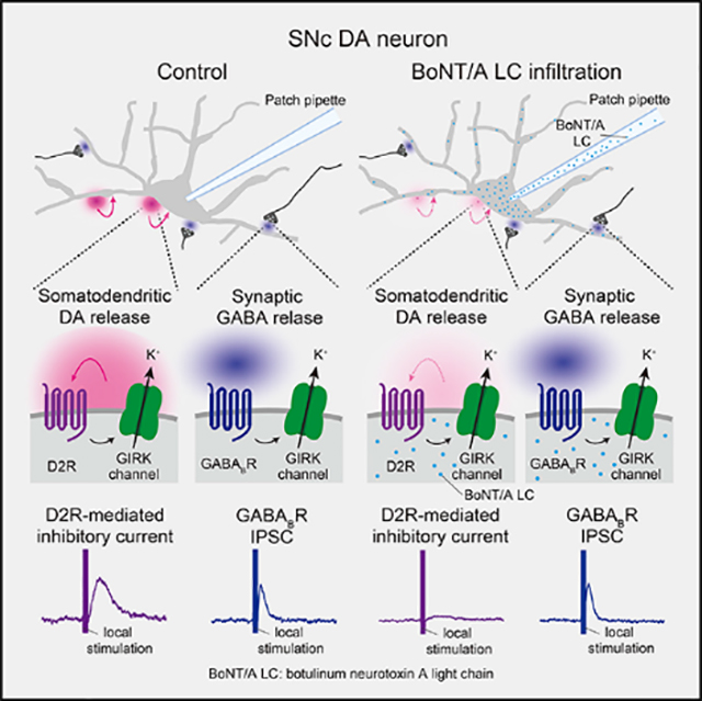
      Bookmark
          There is no author summary for this article yet. Authors can add summaries to their articles on ScienceOpen to make them more accessible to a non-specialist audience.

          SUMMARY

          Somatodendritic dopamine (DA) release from midbrain DA neurons activates D2 autoreceptors on these cells to regulate their activity. However, the source of autoregulatory DA remains controversial. Here, we test the hypothesis that D2 autoreceptors on a given DA neuron in the substantia nigra pars compacta (SNc) are activated primarily by DA released from that same cell, rather than from its neighbors. Voltage-clamp recording allows monitoring of evoked D2-receptor-mediated inhibitory currents (D2ICs) in SNc DA neurons as an index of DA release. Single-cell application of antibodies to Na + channels via the recording pipette decreases spontaneous activity of recorded neurons and attenuates evoked D2ICs; antibodies to SNAP-25, a soluble N-ethylmaleimide-sensitive factor attachment protein receptor (SNARE) protein, also decrease D2IC amplitude. Evoked D2ICs are nearly abolished by the light chain of botulinum neurotoxin A, which cleaves SNAP-25, whereas synaptically activated GABA B-receptor-mediated currents are unaffected. Thus, somatodendritic DA release in the SNc autoinhibits the neuron that releases it.

          In brief

          Somatodendritic dopamine (DA) release inhibits DA neuron activity via D2 autoreceptors. A prevailing hypothesis is that this is mediated by synaptic release from neighboring DA neurons. Hikima et al. show instead that a given SNc DA neuron is regulated by its own released DA, thus exhibiting true autoinhibition.

          Graphical Abstract

          Related collections

          Most cited references94

          • Record: found
          • Abstract: found
          • Article: not found

          Modulation of striatal projection systems by dopamine.

          The basal ganglia are a chain of subcortical nuclei that facilitate action selection. Two striatal projection systems--so-called direct and indirect pathways--form the functional backbone of the basal ganglia circuit. Twenty years ago, investigators proposed that the striatum's ability to use dopamine (DA) rise and fall to control action selection was due to the segregation of D(1) and D(2) DA receptors in direct- and indirect-pathway spiny projection neurons. Although this hypothesis sparked a debate, the evidence that has accumulated since then clearly supports this model. Recent advances in the means of marking neural circuits with optical or molecular reporters have revealed a clear-cut dichotomy between these two cell types at the molecular, anatomical, and physiological levels. The contrast provided by these studies has provided new insights into how the striatum responds to fluctuations in DA signaling and how diseases that alter this signaling change striatal function.
            Bookmark
            • Record: found
            • Abstract: found
            • Article: not found

            The functional anatomy of basal ganglia disorders.

            Basal ganglia disorders are a heterogeneous group of clinical syndromes with a common anatomic locus within the basal ganglia. To account for the variety of clinical manifestations associated with insults to various parts of the basal ganglia we propose a model in which specific types of basal ganglia disorders are associated with changes in the function of subpopulations of striatal projection neurons. This model is based on a synthesis of experimental animal and post-mortem human anatomic and neurochemical data. Hyperkinetic disorders, which are characterized by an excess of abnormal movements, are postulated to result from the selective impairment of striatal neurons projecting to the lateral globus pallidus. Hypokinetic disorders, such as Parkinson's disease, are hypothesized to result from a complex series of changes in the activity of striatal projection neuron subpopulations resulting in an increase in basal ganglia output. This model suggests that the activity of subpopulations of striatal projection neurons is differentially regulated by striatal afferents and that different striatal projection neuron subpopulations may mediate different aspects of motor control.
              Bookmark
              • Record: found
              • Abstract: found
              • Article: not found

              Molecular anatomy of a trafficking organelle.

              Membrane traffic in eukaryotic cells involves transport of vesicles that bud from a donor compartment and fuse with an acceptor compartment. Common principles of budding and fusion have emerged, and many of the proteins involved in these events are now known. However, a detailed picture of an entire trafficking organelle is not yet available. Using synaptic vesicles as a model, we have now determined the protein and lipid composition; measured vesicle size, density, and mass; calculated the average protein and lipid mass per vesicle; and determined the copy number of more than a dozen major constituents. A model has been constructed that integrates all quantitative data and includes structural models of abundant proteins. Synaptic vesicles are dominated by proteins, possess a surprising diversity of trafficking proteins, and, with the exception of the V-ATPase that is present in only one to two copies, contain numerous copies of proteins essential for membrane traffic and neurotransmitter uptake.
                Bookmark

                Author and article information

                Journal
                101573691
                39703
                Cell Rep
                Cell Rep
                Cell reports
                2211-1247
                25 May 2021
                06 April 2021
                09 June 2021
                : 35
                : 1
                : 108951
                Affiliations
                [1 ]Department of Neurosurgery, New York University Grossman School of Medicine, New York, NY 10016, USA
                [2 ]Department of Biochemistry & Molecular Pharmacology, New York University Grossman School of Medicine, New York, NY 10016, USA
                [3 ]Department of Neuroscience & Physiology, New York University Grossman School of Medicine, New York, NY 10016, USA
                [4 ]Lead contact
                Author notes

                AUTHOR CONTRIBUTIONS

                T.H. and M.E.R. designed experiments; T.H. and C.R.L. conducted whole-cell recording experiments; P.W. and J.C conducted immunocytochemical studies; K.I. created and provided BoNT/A light chain, BoNT/A holotoxin, and insight into sample preparation and design of BoNT/A controls; T.H., P.W., and M.E.R. wrote the manuscript with input from all other authors.

                [* ]Correspondence: margaret.rice@ 123456nyu.edu
                Article
                NIHMS1704196
                10.1016/j.celrep.2021.108951
                8189326
                33826884
                482e5088-b1fb-4479-9345-91c68e15f1bc

                This is an open access article under the CC BY-NC-ND license ( http://creativecommons.org/licenses/by-nc-nd/4.0/).

                History
                Categories
                Article

                Cell biology
                Cell biology

                Comments

                Comment on this article