9
views
0
recommends
+1 Recommend
0 collections
    0
    shares
      • Record: found
      • Abstract: found
      • Article: found
      Is Open Access

      Western Immunoblotting for the Diagnosis of Enterococcus faecalis and Streptococcus gallolyticus Infective Endocarditis

      research-article

      Read this article at

      Bookmark
          There is no author summary for this article yet. Authors can add summaries to their articles on ScienceOpen to make them more accessible to a non-specialist audience.

          Abstract

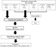
          Blood culture-negative endocarditis (BCNE) remains a diagnostic challenge. In our center, despite a systematic and exhaustive microbiological diagnostics strategy, 22% of patients with BCNE remain without an identified etiology. In an effort to determine the relevance of using Western blot (WB) for the etiological diagnosis of BCNE in patients with early antibiotic use, we developed specific assays for the major infective endocarditis (IE) causative agents, namely, Staphylococcus aureus, Enterococcus faecalis, Streptococcus anginosus, and Streptococcus gallolyticus. Our technique was effective to identify the antigenic profiles of the four tested agents, but cross-reactions with S. aureus and S. anginosus antigens were frequent. A scoring method was developed for the diagnosis of E. faecalis and S. gallolyticus IE using the presence of reactivity to at least two antigenic bands for each bacterium and the positivity to at least one of the Ef300, Ef72, or Ef36 proteic bands for E. faecalis, and positivity for the two Sg75 and Sg97 proteic bands for S. gallolyticus. We tested these diagnostic criteria in a prospective cohort of 363 patients with suspected IE. Immunoblotting for the diagnosis of E. faecalis IE showed a sensitivity of 100% and a specificity of 99%. The positive and negative predictive values were 73 and 100%, respectively. Regarding S. gallolyticus infection, immunoblot had a sensitivity of 100% and a specificity of 95%. However, the positive predictive value was 22%, whereas the predictive negative value was 100%. Using WB, we identified a potential etiological agent in 4 of 14 BCNE cases with no identified pathogen. In conclusion, WB constitutes a promising and helpful method to diagnose E. faecalis or S. gallolyticus IE in patients with early antibiotic uptake and negative blood cultures.

          Related collections

          Most cited references9

          • Record: found
          • Abstract: found
          • Article: found
          Is Open Access

          Blood culture-negative endocarditis

          Supplemental Digital Content is available in the text
            Bookmark
            • Record: found
            • Abstract: found
            • Article: not found

            Serological cross-reactions between Bartonella and Chlamydia species: implications for diagnosis.

            Diagnosis of Chlamydia or Bartonella infections continues to rely mainly on serology. However, serological cross-reactions between members of these genera have recently been described. Sera from eight patients originally diagnosed as having Chlamydia pneumoniae endocarditis reacted with both Chlamydia sp. and Bartonella quintana antigens (microimmunofluorescence technique). Adsorption of sera with B. quintana or C. pneumoniae antigens removed anti-C. pneumoniae antibodies, whereas adsorption with C. pneumoniae antigens did not change antibody titers to B. quintana. Western blot analysis confirmed the presence of cross-reacting antigens and showed antibody patterns in all sera to be compatible with a Bartonella infection. These patients were therefore probably suffering from Bartonella-induced rather than Chlamydia-induced endocarditis. In contrast, sera from 10 patients presumed to be suffering from C. pneumoniae pneumonia did not display anti-B. quintana antibodies, although cross-reacting antigens were revealed by Western blotting. This work highlights the possibility that cases of infective Bartonella endocarditis are erroneously diagnosed as chlamydial infections.
              Bookmark
              • Record: found
              • Abstract: found
              • Article: not found

              Differences between endocarditis caused by Streptococcus bovis and Enterococcus spp. and their association with colorectal cancer.

              Streptococcus bovis group and Enterococcus spp. share phenotypic characteristics and intestinal habitat. Both have been associated with endocarditis and colorectal neoplasm (CRN). We studied all cases of endocarditis diagnosed between 1988 and 2014 in our centre and caused by S. bovis (109, 48.8 % of the bacteremia) and by Enterococcus spp. (36, 3.4 % of the bacteremia). Patients were seen until death or during a long-term follow-up, in order to rule out a concomitant CRN. The 109 cases of S. bovis endocarditis (SbIE) compared with the 36 caused by enterococci showed: a higher proportion of males (91 % vs. 72 %, p=0.005), more multivalvular involvement (28 % vs. 6 %, p=0.004), embolic complications (44 vs. 22 %, p=0.02) and colorectal neoplasm (64 % vs. 25 %, p=0.001). SbIE showed fewer co-morbidities (32 vs. 58 %, p=0.005), and less frequently urinary infection source (0 vs. 25 %, p=0.001) and healthcare-related infection (2 vs. 44 %, p=0.001). A total of 123 patients were followed up for an extended period (mean: 65.9 ± 57.5 months). During the follow-up, 6 of 28 (21 %) cases with enterococcal endocarditis and 43 of 95 (45.2 %, p=0.01) cases with SbIE developed a new CRN. These neoplasiae appeared a mean of 60.4 months later (range 12-181 months). Among the 43 cases with SbIE and CRN, 12 had had a previously normal colonoscopy and 31 had had a previous CRN and developed a second neoplasm. Cases of SbIE present important differences with those caused by Enterococcus spp. Colonoscopy must be mandatory both in the initial evaluation of SbIE, as during the follow-up period.
                Bookmark

                Author and article information

                Contributors
                Journal
                Front Cell Infect Microbiol
                Front Cell Infect Microbiol
                Front. Cell. Infect. Microbiol.
                Frontiers in Cellular and Infection Microbiology
                Frontiers Media S.A.
                2235-2988
                12 September 2019
                2019
                : 9
                : 314
                Affiliations
                [1] 1Aix Marseille Univ., IRD, AP-HM, MEPHI , Marseille, France
                [2] 2Service de Cardiologie, Hôpital de la Timone , Marseille, France
                [3] 3Microbiology Laboratory, Institut Hospitalo-Universitaire (IHU) Mediterranée Infection , Marseille, France
                [4] 4Aix Marseille Univ., IRD, AP-HM, SSA, VITROME, IHU Méditerranée Infection , Marseille, France
                [5] 5CNR des Rickettsies, fièvre Q, Bartonella, IHU Méditerranée Infection , Marseille, France
                Author notes

                Edited by: Max Maurin, Université Grenoble Alpes, France

                Reviewed by: Cristiane Da Cruz Lamas, Instituto Nacional de Cardiologia, Brazil; Juan M. Pericas, University Hospital Arnau de Vilanova, Spain

                *Correspondence: Didier Raoult didier.raoult@ 123456gmail.com

                This article was submitted to Clinical Microbiology, a section of the journal Frontiers in Cellular and Infection Microbiology

                †These authors have contributed equally to this work

                Article
                10.3389/fcimb.2019.00314
                6751308
                31572688
                46cb8a0a-8573-4458-a369-9bf8daa4dec9
                Copyright © 2019 Arregle, Gouriet, Amphoux, Edouard, Chaudet, Casalta, Habib, Fournier and Raoult.

                This is an open-access article distributed under the terms of the Creative Commons Attribution License (CC BY). The use, distribution or reproduction in other forums is permitted, provided the original author(s) and the copyright owner(s) are credited and that the original publication in this journal is cited, in accordance with accepted academic practice. No use, distribution or reproduction is permitted which does not comply with these terms.

                History
                : 24 June 2019
                : 21 August 2019
                Page count
                Figures: 4, Tables: 2, Equations: 0, References: 15, Pages: 7, Words: 4885
                Categories
                Cellular and Infection Microbiology
                Original Research

                Infectious disease & Microbiology
                blood culture negative endocarditis,enterococcus faecalis,streptococcus gallolyticus,western immunoblotting,serology

                Comments

                Comment on this article