29
views
0
recommends
+1 Recommend
0 collections
    0
    shares
      • Record: found
      • Abstract: found
      • Article: not found

      Role of YidC in folding of polytopic membrane proteins

      research-article

      Read this article at

      ScienceOpenPublisherPMC
      Bookmark
          There is no author summary for this article yet. Authors can add summaries to their articles on ScienceOpen to make them more accessible to a non-specialist audience.

          Abstract

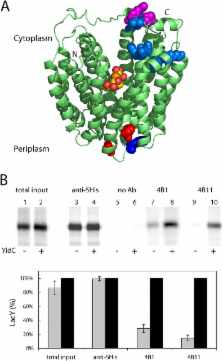
          YidC of Echerichia coli, a member of the conserved Alb3/Oxa1/YidC family, is postulated to be important for biogenesis of membrane proteins. Here, we use as a model the lactose permease (LacY), a membrane transport protein with a known three-dimensional structure, to determine whether YidC plays a role in polytopic membrane protein insertion and/or folding. Experiments in vivo and with an in vitro transcription/translation/insertion system demonstrate that YidC is not necessary for insertion per se, but plays an important role in folding of LacY. By using the in vitro system and two monoclonal antibodies directed against conformational epitopes, LacY is shown to bind the antibodies poorly in YidC-depleted membranes. Moreover, LacY also folds improperly in proteoliposomes prepared without YidC. However, when the proteoliposomes are supplemented with purified YidC, LacY folds correctly. The results indicate that YidC plays a primary role in folding of LacY into its final tertiary conformation via an interaction that likely occurs transiently during insertion into the lipid phase of the membrane.

          Related collections

          Most cited references54

          • Record: found
          • Abstract: found
          • Article: not found

          X-ray structure of a protein-conducting channel.

          A conserved heterotrimeric membrane protein complex, the Sec61 or SecY complex, forms a protein-conducting channel, allowing polypeptides to be transferred across or integrated into membranes. We report the crystal structure of the complex from Methanococcus jannaschii at a resolution of 3.2 A. The structure suggests that one copy of the heterotrimer serves as a functional translocation channel. The alpha-subunit has two linked halves, transmembrane segments 1-5 and 6-10, clamped together by the gamma-subunit. A cytoplasmic funnel leading into the channel is plugged by a short helix. Plug displacement can open the channel into an 'hourglass' with a ring of hydrophobic residues at its constriction. This ring may form a seal around the translocating polypeptide, hindering the permeation of other molecules. The structure also suggests mechanisms for signal-sequence recognition and for the lateral exit of transmembrane segments of nascent membrane proteins into lipid, and indicates binding sites for partners that provide the driving force for translocation.
            Bookmark
            • Record: found
            • Abstract: found
            • Article: not found

            YidC mediates membrane protein insertion in bacteria.

            The basic machinery for the translocation of proteins into or across membranes is remarkably conserved from Escherichia coli to humans. In eukaryotes, proteins are inserted into the endoplasmic reticulum using the signal recognition particle (SRP) and the SRP receptor, as well as the integral membrane Sec61 trimeric complex (composed of alpha, beta and gamma subunits). In bacteria, most proteins are inserted by a related pathway that includes the SRP homologue Ffh, the SRP receptor FtsY, and the SecYEG trimeric complex, where Y and E are related to the Sec61 alpha and gamma subunits, respectively. Proteins in bacteria that exhibit no dependence on the Sec translocase were previously thought to insert into the membrane directly without the aid of a protein machinery. Here we show that membrane insertion of two Sec-independent proteins requires YidC. YidC is essential for E. coli viability and homologues are present in mitochondria and chloroplasts. Depletion of YidC also interferes with insertion of Sec-dependent membrane proteins, but it has only a minor effect on the export of secretory proteins. These results provide evidence for an additional component of the translocation machinery that is specialized for the integration of membrane proteins.
              Bookmark
              • Record: found
              • Abstract: found
              • Article: not found

              YidC, the Escherichia coli homologue of mitochondrial Oxa1p, is a component of the Sec translocase.

              In Escherichia coli, both secretory and inner membrane proteins initially are targeted to the core SecYEG inner membrane translocase. Previous work has also identified the peripherally associated SecA protein as well as the SecD, SecF and YajC inner membrane proteins as components of the translocase. Here, we use a cross-linking approach to show that hydrophilic portions of a co-translationally targeted inner membrane protein (FtsQ) are close to SecA and SecY, suggesting that insertion takes place at the SecA/Y interface. The hydrophobic FtsQ signal anchor sequence contacts both lipids and a novel 60 kDa translocase-associated component that we identify as YidC. YidC is homologous to Saccharomyces cerevisiae Oxa1p, which has been shown to function in a novel export pathway at the mitochondrial inner membrane. We propose that YidC is involved in the insertion of hydrophobic sequences into the lipid bilayer after initial recognition by the SecAYEG translocase.
                Bookmark

                Author and article information

                Journal
                J Cell Biol
                The Journal of Cell Biology
                The Rockefeller University Press
                0021-9525
                1540-8140
                12 April 2004
                : 165
                : 1
                : 53-62
                Affiliations
                [1 ]Howard Hughes Medical Institute, Immunology, and Molecular Genetics, Molecular Biology Institute, University of California, Los Angeles, Los Angeles, CA 90095
                [2 ]Department of Physiology, Immunology, and Molecular Genetics, Molecular Biology Institute, University of California, Los Angeles, Los Angeles, CA 90095
                [3 ]Department of Microbiology, Immunology, and Molecular Genetics, Molecular Biology Institute, University of California, Los Angeles, Los Angeles, CA 90095
                Author notes

                Address correspondence to H. Ronald Kaback, 5-748 Macdonald Research Laboratories, Rm. 6720, P.O. Box 951662, Howard Hughes Medical Institute, University of California, Los Angeles, Los Angeles, CA 90095-1662. Tel.: (310) 206-5053. Fax: (310) 206-8623. email: RonaldK@ 123456hhmi.ucla.edu

                Article
                200402067
                10.1083/jcb.200402067
                2172087
                15067017
                422a2c4b-7d6b-498d-8ec0-90512dcd1bb5
                Copyright © 2004, The Rockefeller University Press
                History
                : 12 February 2004
                : 9 March 2004
                Categories
                Article

                Cell biology
                lacy; membrane insertion; protein folding; secyeg; oxa1/alb3 protein family

                Comments

                Comment on this article