13
views
0
recommends
+1 Recommend
0 collections
    0
    shares
      • Record: found
      • Abstract: found
      • Article: found
      Is Open Access

      Diffusion tensor imaging and tractwise fractional anisotropy statistics: quantitative analysis in white matter pathology

      research-article

      Read this article at

      Bookmark
          There is no author summary for this article yet. Authors can add summaries to their articles on ScienceOpen to make them more accessible to a non-specialist audience.

          Abstract

          Background

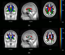
          Information on anatomical connectivity in the brain by measurements of the diffusion of water in white matter tracts lead to quantification of local tract directionality and integrity.

          Methods

          The combination of connectivity mapping (fibre tracking, FT) with quantitative diffusion fractional anisotropy (FA) mapping resulted in the approach of results based on group-averaged data, named tractwise FA statistics (TFAS). The task of this study was to apply these methods to group-averaged data from different subjects to quantify differences between normal subjects and subjects with defined alterations of the corpus callosum (CC).

          Results

          TFAS exhibited a significant FA reduction especially in the CC, in agreement with region of interest (ROI)-based analyses.

          Conclusion

          In summary, the applicability of the TFAS approach to diffusion tensor imaging studies of normal and pathologically altered brains was demonstrated.

          Related collections

          Most cited references27

          • Record: found
          • Abstract: not found
          • Article: not found

          Spatial registration and normalization of images

            Bookmark
            • Record: found
            • Abstract: not found
            • Article: not found

            Automatic 3D Intersubject Registration of MR Volumetric Data in Standardized Talairach Space

              Bookmark
              • Record: found
              • Abstract: found
              • Article: not found

              Automatic 3D intersubject registration of MR volumetric data in standardized Talairach space.

              In both diagnostic and research applications, the interpretation of MR images of the human brain is facilitated when different data sets can be compared by visual inspection of equivalent anatomical planes. Quantitative analysis with predefined atlas templates often requires the initial alignment of atlas and image planes. Unfortunately, the axial planes acquired during separate scanning sessions are often different in their relative position and orientation, and these slices are not coplanar with those in the atlas. We have developed a completely automatic method to register a given volumetric data set with Talairach stereotaxic coordinate system. The registration method is based on multi-scale, three-dimensional (3D) cross-correlation with an average (n > 300) MR brain image volume aligned with the Talariach stereotaxic space. Once the data set is re-sampled by the transformation recovered by the algorithm, atlas slices can be directly superimposed on the corresponding slices of the re-sampled volume. the use of such a standardized space also allows the direct comparison, voxel to voxel, of two or more data sets brought into stereotaxic space. With use of a two-tailed Student t test for paired samples, there was no significant difference in the transformation parameters recovered by the automatic algorithm when compared with two manual landmark-based methods (p > 0.1 for all parameters except y-scale, where p > 0.05). Using root-mean-square difference between normalized voxel intensities as an unbiased measure of registration, we show that when estimated and averaged over 60 volumetric MR images in standard space, this measure was 30% lower for the automatic technique than the manual method, indicating better registrations. Likewise, the automatic method showed a 57% reduction in standard deviation, implying a more stable technique. The algorithm is able to recover the transformation even when data are missing from the top or bottom of the volume. We present a fully automatic registration method to map volumetric data into stereotaxic space that yields results comparable with those of manually based techniques. The method requires no manual identification of points or contours and therefore does not suffer the drawbacks involved in user intervention such as reproducibility and interobserver variability.
                Bookmark

                Author and article information

                Journal
                Biomed Eng Online
                BioMedical Engineering OnLine
                BioMed Central
                1475-925X
                2007
                9 November 2007
                : 6
                : 42
                Affiliations
                [1 ]Department of Neurology, University of Ulm, Ulm, Germany
                Article
                1475-925X-6-42
                10.1186/1475-925X-6-42
                2186341
                17996104
                421f47f9-459a-436c-b236-7ccd810ef0ef
                Copyright © 2007 Mueller et al; licensee BioMed Central Ltd.

                This is an Open Access article distributed under the terms of the Creative Commons Attribution License ( http://creativecommons.org/licenses/by/2.0), which permits unrestricted use, distribution, and reproduction in any medium, provided the original work is properly cited.

                History
                : 2 July 2007
                : 9 November 2007
                Categories
                Research

                Biomedical engineering
                Biomedical engineering

                Comments

                Comment on this article