0
views
0
recommends
+1 Recommend
0 collections
    0
    shares
      • Record: found
      • Abstract: found
      • Article: found
      Is Open Access

      Fast progressive lower motor neuron disease is an ALS variant: A two-centre tract of interest-based MRI data analysis

      research-article

      Read this article at

      Bookmark
          There is no author summary for this article yet. Authors can add summaries to their articles on ScienceOpen to make them more accessible to a non-specialist audience.

          Abstract

          Background

          The criteria for assessing upper motor neuron pathology in pure lower motor neuron disease (LMND) still remain a major issue of debate with respect to the clinical classification as an amyotrophic lateral sclerosis (ALS) variant.

          Objective

          The study was designed to investigate white matter damage by a hypothesis-guided tract-of-interest-based approach in patients with LMND compared with healthy controls and ´classical´ ALS patients in order to identify in vivo brain structural changes according to the neuropathologically defined ALS affectation pattern. Data were pooled from two previous studies at two different study sites (Ulm, Germany and Milano, Italy).

          Methods

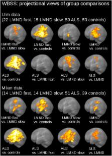
          DTI-based white matter integrity mapping was performed by voxelwise statistical comparison and by a tractwise analysis of fractional anisotropy (FA) maps according to the ALS-staging pattern for 65 LMND patients (clinically differentiated in fast and slow progressors) vs. 92 matched controls and 101 ALS patients with a ‘classical’ phenotype to identify white matter structural alterations.

          Results

          The analysis of white matter structural connectivity by regional FA reductions demonstrated the characteristic alteration patterns along the CST and also in frontal and prefrontal brain areas in LMND patients compared to controls and ALS. Fast progressing LMND showed substantial involvement, like in ALS, while slow progressors showed less severe alterations. In the tract-specific analysis according to the ALS-staging pattern, fast progressing LMND showed significant alterations of ALS-related tract systems as compared to slow progressors and controls.

          Conclusions

          This study showed an affectation pattern for corticoefferent fibers in LMND with fast disease progression as defined for ALS, that way confirming the hypothesis that fast progressing LMND is a phenotypical variant of ALS.

          Highlights

          • LMND is associated with brain alterations along the CST and in frontal areas.

          • Fast progressive LMND show cerebral tract involvement like in ALS.

          • DTI supports that fast progressive LMND is a phenotypical variant of ALS.

          Related collections

          Most cited references22

          • Record: found
          • Abstract: found
          • Article: not found

          Phenotypic heterogeneity of amyotrophic lateral sclerosis: a population based study.

          Different amyotrophic lateral sclerosis (ALS) phenotypes have been recognised, marked by a varying involvement of spinal and bulbar upper and lower motor neurons. However, the differential characteristics of these phenotypes are still largely unknown. To define the epidemiology and outcome of ALS phenotypes in a population based setting. All ALS cases incident in two Italian regions were prospectively collected from 1995 to 2004 in an epidemiological register. Cases were classified according to established ALS phenotypes: classic, bulbar, flail arm, flail leg, pyramidal, respiratory, pure lower motor neuron (PLMN) and pure upper motor neuron (PUMN). ALS phenotype were determined in 1332 out of 1351 incident patients (98.6%). Classic and bulbar phenotypes had similar mean annual incidence rates. Gender specific incidence rates showed a male preponderance in respiratory, flail arm, classic and PLMN phenotypes; in all other phenotypes, men and women had similar incidence rates. Age at onset was significantly lower in pyramidal, PLMN and PUMN phenotypes and higher in the bulbar phenotype. The best outcomes were observed in PUMN, pyramidal, PLMN and flail arm phenotypes and the worst in respiratory and bulbar phenotypes. Our epidemiological findings suggest that ALS phenotypes carry distinctive and easily distinguishable clinical and prognostic characteristics, strongly related to a complex interplay between gender and age. The categorisation of ALS patients according to more homogenous clinical groups is relevant in identifying biological markers for ALS and should be considered for the design of clinical trials.
            Bookmark
            • Record: found
            • Abstract: found
            • Article: not found

            The effect of filter size on VBM analyses of DT-MRI data.

            Voxel-based morphometry (VBM) has been used to analyze diffusion tensor MRI (DT-MRI) data in a number of studies. In VBM, following spatial normalization, data are smoothed to improve the validity of statistical inferences and to reduce inter-individual variation. However, the size of the smoothing filter used for VBM of DT-MRI data is highly variable across studies. For example, a literature review revealed that Gaussian smoothing kernels ranging in size (full width at half maximum) from zero to 16 mm have been used in DT-MRI VBM type studies. To investigate the effect of varying filter size in such analyses, whole brain DT-MRI data from 14 schizophrenic patients were compared with those of 14 matched control subjects using VBM, when the filter size was varied from zero to 16 mm. Within this range of smoothing, four different conclusions regarding apparent patient control differences could be made: (i) no significant patient-control differences; (ii) reduced FA in right superior temporal gyrus (STG) in patients; (iii) reduced FA in both right STG and left cerebellum in patients; and (iv) reduced FA only in left cerebellum in patients. These findings stress the importance of recognizing the effect of the matched filter theorem on VBM analyses of DT-MRI data. Finally, we investigated whether one of the underlying assumptions of parametric VBM, i.e., the normality of the residuals, is met. Our results suggest that, even with moderate smoothing, a large number of voxels within central white matter regions may have non-normally distributed residuals thus making valid statistical inferences with a parametric approach problematic in these areas.
              Bookmark
              • Record: found
              • Abstract: found
              • Article: not found

              Diffusion tensor imaging analysis of sequential spreading of disease in amyotrophic lateral sclerosis confirms patterns of TDP-43 pathology.

              Diffusion tensor imaging can identify amyotrophic lateral sclerosis-associated patterns of brain alterations at the group level. Recently, a neuropathological staging system for amyotrophic lateral sclerosis has shown that amyotrophic lateral sclerosis may disseminate in a sequential regional pattern during four disease stages. The objective of the present study was to apply a new methodological diffusion tensor imaging-based approach to automatically analyse in vivo the fibre tracts that are prone to be involved at each neuropathological stage of amyotrophic lateral sclerosis. Two data samples, consisting of 130 diffusion tensor imaging data sets acquired at 1.5 T from 78 patients with amyotrophic lateral sclerosis and 52 control subjects; and 55 diffusion-tensor imaging data sets at 3.0 T from 33 patients with amyotrophic lateral sclerosis and 22 control subjects, were analysed by a tract of interest-based fibre tracking approach to analyse five tracts that become involved during the course of amyotrophic lateral sclerosis: the corticospinal tract (stage 1); the corticorubral and the corticopontine tracts (stage 2); the corticostriatal pathway (stage 3); the proximal portion of the perforant path (stage 4); and two reference pathways. The statistical analyses of tracts of interest showed differences between patients with amyotrophic lateral sclerosis and control subjects for all tracts. The significance level of the comparisons at the group level was lower, the higher the disease stage with corresponding involved fibre tracts. Both the clinical phenotype as assessed by the amyotrophic lateral sclerosis functional rating scale-revised and disease duration correlated significantly with the resulting staging scheme. In summary, the tract of interest-based technique allowed for individual analysis of predefined tract structures, thus making it possible to image in vivo the disease stages in amyotrophic lateral sclerosis. This approach can be used not only for individual clinical work-up purposes, but enlarges the spectrum of potential non-invasive surrogate markers as a neuroimaging-based read-out for amyotrophic lateral sclerosis studies within a clinical context. © The Author (2014). Published by Oxford University Press on behalf of the Guarantors of Brain. All rights reserved. For Permissions, please email: journals.permissions@oup.com.
                Bookmark

                Author and article information

                Contributors
                Journal
                Neuroimage Clin
                Neuroimage Clin
                NeuroImage : Clinical
                Elsevier
                2213-1582
                14 October 2017
                2018
                14 October 2017
                : 17
                : 145-152
                Affiliations
                [a ]Department of Neurology, University of Ulm, Germany
                [b ]Neuroimaging Research Unit, Institute of Experimental Neurology, Division of Neuroscience, San Raffaele Scientific Institute, Vita-Salute San Raffaele University, Milan, Italy
                [c ]Department of Neurology, Institute of Experimental Neurology, Division of Neuroscience, San Raffaele Scientific Institute, Milan, Italy
                Author notes
                [* ]Corresponding author at: Dept. of Neurology, University of Ulm, Oberer Eselsberg 45, 89081 Ulm, Germany.Dept. of NeurologyUniversity of UlmOberer Eselsberg 45Ulm89081Germany jan.kassubek@ 123456uni-ulm.de
                [1]

                Shared first authorship.

                [2]

                Shared senior authorship.

                Article
                S2213-1582(17)30249-8
                10.1016/j.nicl.2017.10.008
                5651542
                29071208
                b5b736d5-c75d-41f9-bae5-56e39493ae54
                © 2017 The Authors

                This is an open access article under the CC BY-NC-ND license (http://creativecommons.org/licenses/by-nc-nd/4.0/).

                History
                : 20 July 2017
                : 7 September 2017
                : 6 October 2017
                Categories
                Regular Article

                dti,lower motor neuron disease,als,pma,mri
                dti, lower motor neuron disease, als, pma, mri

                Comments

                Comment on this article