117
views
0
recommends
+1 Recommend
0 collections
    0
    shares
      • Record: found
      • Abstract: found
      • Article: found
      Is Open Access

      Perianal Median Raphe Cyst: A Rare Lesion with Unusual Histology and Localization

      case-report

      Read this article at

      Bookmark
          There is no author summary for this article yet. Authors can add summaries to their articles on ScienceOpen to make them more accessible to a non-specialist audience.

          Abstract

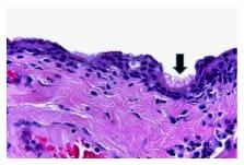
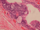
          Median raphe cysts present anywhere between the external urethral meatus and the anus. The cysts can occur at parameatus, glans penis, penile shaft, scrotum, or perineum. Perianal region is an extremely rare location for these lesions. Here we present a 50-year-old male patient who presented with a cystic, fluctuant lesion, located at 12 o'clock in perianal region. Microscopic examination revealed a cystic lesion with keratinized and nonkeratinized stratified squamous epithelium, pseudostratified ciliated epithelium, and scattered goblet cells. The final diagnosis of the lesion was median raphe cyst. Ciliated cells and perianal localization in median raphe cysts are extremely rare characteristics.

          Related collections

          Most cited references12

          • Record: found
          • Abstract: found
          • Article: found
          Is Open Access

          Male median raphe cysts: serial retrospective analysis and histopathological classification

          Background To review the clinical and pathological characteristics of median raphe cysts and to classify the lesions according to pathogenesis and histopathological findings. Methods The medical records of patients who were diagnosed with median raphe cysts between 2001 and 2010 were reviewed to document the clinical presentation and pathological findings of the cysts. Results Most patients were asymptomatic; however, 9 patients had inflammatory or infectious cysts that were tender or painful. Four patients who had cysts on the parameatus and distal prepuce had difficulty voiding. Hematuria and hematospermia were noted in 2 cases. Thirty-one cysts were lined with an urothelium-like epithelium, and a squamous epithelium lining was found in 3 cases. In 2 cases, a well-formed mucinous glandular structure was observed. The other 20 cysts consisted of mixed epithelia. After excision of the cysts under local or general anesthesia, an urethral fistula developed as a complication in only 1 case. Conclusions Median raphe cysts are benign lesions formed due to tissue trapping during the development of urethral folds. The cysts can be defined into 4 types based on pathological findings: urethral, epidermoid, glandular, and mixed. The associated symptoms and signs should be taken into consideration when determining the treatment for the cysts. Virtual slides The virtual slide(s) for this article can be found here: http//http://www.diagnosticpathology.diagnomx.eu/vs/7727074877500751
            Bookmark
            • Record: found
            • Abstract: found
            • Article: not found

            Median raphe (parameatal) cysts of the penis.

            Although median raphe cysts of the penis have been widely reported the pathogenesis is obscure. We describe the microstructural detection of 3 cases of median raphe to study the true pathogenesis. Three patients presented to our clinic of plastic surgery complaining of abnormal shape of the penis or mass recurrence. High magnification light microscopy and electron microscopy were performed. Microstructural study revealed that all 3 cases had similar findings to those of the common urethral mucosa. Median raphe cysts are derived from the external paraurethral ducts and contain features that to our knowledge have not been previously reported.
              Bookmark
              • Record: found
              • Abstract: not found
              • Article: not found

              Median raphe cyst on the scrotum and perineum.

                Bookmark

                Author and article information

                Journal
                Case Rep Dermatol Med
                Case Rep Dermatol Med
                CRIDM
                Case Reports in Dermatological Medicine
                Hindawi Publishing Corporation
                2090-6463
                2090-6471
                2015
                22 February 2015
                : 2015
                : 487814
                Affiliations
                School of Medicine, Department of Pathology, Akdeniz University, 07070 Antalya, Turkey
                Author notes

                Academic Editor: Akimichi Morita

                Article
                10.1155/2015/487814
                4352436
                41e16aee-2d15-418a-8a90-0537c648acfe
                Copyright © 2015 Betül Ünal et al.

                This is an open access article distributed under the Creative Commons Attribution License, which permits unrestricted use, distribution, and reproduction in any medium, provided the original work is properly cited.

                History
                : 25 November 2014
                : 25 January 2015
                Categories
                Case Report

                Dermatology
                Dermatology

                Comments

                Comment on this article