16
views
0
recommends
+1 Recommend
0 collections
    0
    shares
      • Record: found
      • Abstract: found
      • Article: found
      Is Open Access

      Unusual remodeling of the hyalinization band in vulval lichen sclerosus by type V collagen and ECM 1 protein

      research-article

      Read this article at

      Bookmark
          There is no author summary for this article yet. Authors can add summaries to their articles on ScienceOpen to make them more accessible to a non-specialist audience.

          Abstract

          OBJECTIVES:

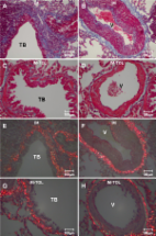
          The vulva is the primary site affected in lichen sclerosus, a chronic dermatosis in women that is histologically characterized by a zone of collagen remodeling in the superior dermis. The normal physiological properties of the vulva depend on the assembly of collagen types I (COLI), III (COLIII) and V (COLV), which form heterotypic fibers, and extracellular matrix protein interactions. COLV regulates the heterotypic fiber diameter, and the preservation of its properties is important for maintaining normal tissue architecture and function. In the current work, we analyzed the expression of COLV and its relationship with COLI, COLIII, elastic fibers and extracellular matrix protein 1 in vulvar biopsies from patients with lichen sclerosus.

          METHODS:

          Skin biopsies from 21 patients with lichen sclerosus, classified according to Hewitt histological criteria, were studied and compared to clinically normal vulvar tissue (N=21). Morphology, immunohistochemistry, immunofluorescence, 3D reconstruction and morphometric analysis of COLI, COLIII, COLV deposition, elastic fibers and extracellular matrix 1 expression in a zone of collagen remodeling in the superior dermis were performed.

          RESULTS:

          A significant decrease of elastic fibers and extracellular matrix 1 protein was present in the hyalinization zone of lichen sclerosus compared to healthy controls. The non-homogeneous distribution of collagen fibers visualized under immunofluorescence in the hyalinization zone of lichen sclerosus and control skin was confirmed by histomorphometry. Lichen sclerosus dermis shows a significant increase of COLI, COLIII and COLV expression compared to the healthy controls. Significant inverse associations were found between elastic fibers and COLV and between COLV and extracellular matrix 1 expression. A direct association was found between elastic fiber content and extracellular matrix 1 expression. Tridimensional reconstruction of the heterotypic fibers of the lichen sclerosus zone of collagen remodeling confirmed the presence of densely clustered COLV.

          CONCLUSIONS:

          Increased deposition of abnormal COLV and its correlation with extracellular matrix 1 and elastic fibers suggest that COLV may be a trigger in the pathogenesis of lichen sclerosus.

          Related collections

          Most cited references102

          • Record: found
          • Abstract: found
          • Article: not found

          Collagen fibrillogenesis in vitro: interaction of types I and V collagen regulates fibril diameter.

          The small-diameter fibrils of the chick corneal stroma are heterotypic, composed of both collagen types I and V. This tissue has a high concentration of type V collagen relative to other type I-containing tissues with larger-diameter fibrils, suggesting that heterotypic interactions may have a regulatory role in the control of fibril diameter. The interactions of collagen types I and V were studied using an in vitro self-assembly system. Collagens were purified from lathyritic chick embryos in the presence of protease inhibitors. The type V collagen preparations contained higher molecular weight forms of the alpha 1(V) and alpha 2(V) chains constituting 60-70% of the total. Rotary-shadow electron micrographs showed a persistence of a small, pepsin-sensitive terminal region in an amount consistent with that seen by electrophoresis. In vitro, this purified type V collagen formed thin fibrils with no apparent periodicity, while type I collagen fibrils had a broad distribution of large diameters. However, when type I collagen was mixed with increasing amounts of type V collagen a progressive and significant decrease in both the mean fibril diameter and the variance was observed for D periodic fibrils. The amino-terminal domain of the type V collagen molecule was required for this regulatory effect and in its absence little diameter reducing activity was observed. Electron microscopy using collagen type-specific monoclonal antibodies demonstrated that the fibrils formed were heterotypic, containing both collagen types I and V. These data indicate that the interaction of type V with type I collagen is one mechanism modulating fibril diameter and is at least partially responsible for the regulation of collagen fibril formation.
            Bookmark
            • Record: found
            • Abstract: found
            • Article: not found

            Type V collagen: heterotypic type I/V collagen interactions in the regulation of fibril assembly.

            D Birk (2001)
            Type V collagen is a quantitatively minor fibrillar collagen with a broad tissue distribution. The most common type V collagen isoform is alpha1(V)(2) alpha2(V) found in cornea. However, other isoforms exist, including an [alpha1(V)alpha2(V)alpha3(V)] form, an alpha1(V)(3) homotrimer and hybrid type V/XI forms. The functional role and fibrillar organization of these isoforms is not understood. In the cornea, type V collagen has a key role in the regulation of initial fibril assembly. Type I and type V collagen co-assemble into heterotypic fibrils. The entire triple-helical domain of the type V collagen molecules is buried within the fibril and type I collagen molecules are present along the fibril surface. The retained NH(2)-terminal domains of the type V collagen are exposed at the surface, extending outward through the gap zones. The molecular model of the NH(2)-terminal domain indicates that the short alpha helical region is a flexible hinge-like region allowing the peptide to project away from the major axis of the molecule; the short triple-helical regions serve as an extension through the hole zone, placing the tyrosine-rich domain at the surface. The assembly of early, immature fibril intermediates (segments) is regulated by the NH(2)-terminal domain of type V collagen. These NH(2)-terminal domains alter accretion of collagen molecules onto fibrils and therefore lateral growth. A critical density would favor the initiation of new fibrils rather than the continued growth of existing fibrils. Other type V collagen isoforms are likely to have an important role in non-cornea tissues. This role may be mediated by supramolecular aggregates different from those in the corneal stroma or by an alteration of the interactions mediated by tissue-specific type V collagen domains generated by different isoforms or aggregate structures. Presumably, the aggregate structure or specific domains are involved in the regionalization of fibril-associated macromolecules necessary for the tissue-specific regulation of later fibril growth and matrix assembly stages.
              Bookmark
              • Record: found
              • Abstract: found
              • Article: found
              Is Open Access

              Collagen V-induced nasal tolerance downregulates pulmonary collagen mRNA gene and TGF-beta expression in experimental systemic sclerosis

              Background The purpose of this study was to evaluate collagen deposition, mRNA collagen synthesis and TGF-beta expression in the lung tissue in an experimental model of scleroderma after collagen V-induced nasal tolerance. Methods Female New Zealand rabbits (N = 12) were immunized with 1 mg/ml of collagen V in Freund's adjuvant (IM). After 150 days, six immunized animals were tolerated by nasal administration of collagen V (25 μg/day) (IM-TOL) daily for 60 days. The collagen content was determined by morphometry, and mRNA expressions of types I, III and V collagen were determined by Real-time PCR. The TGF-beta expression was evaluated by immunostaining and quantified by point counting methods. To statistic analysis ANOVA with Bonferroni test were employed for multiple comparison when appropriate and the level of significance was determined to be p < 0.05. Results IM-TOL, when compared to IM, showed significant reduction in total collagen content around the vessels (0.371 ± 0.118 vs. 0.874 ± 0.282, p < 0.001), bronchioles (0.294 ± 0.139 vs. 0.646 ± 0.172, p < 0.001) and in the septal interstitium (0.027 ± 0.014 vs. 0.067 ± 0.039, p = 0.026). The lung tissue of IM-TOL, when compared to IM, showed decreased immunostaining of types I, III and V collagen, reduced mRNA expression of types I (0.10 ± 0.07 vs. 1.0 ± 0.528, p = 0.002) and V (1.12 ± 0.42 vs. 4.74 ± 2.25, p = 0.009) collagen, in addition to decreased TGF-beta expression (p < 0.0001). Conclusions Collagen V-induced nasal tolerance in the experimental model of SSc regulated the pulmonary remodeling process, inhibiting collagen deposition and collagen I and V mRNA synthesis. Additionally, it decreased TGF-beta expression, suggesting a promising therapeutic option for scleroderma treatment.
                Bookmark

                Author and article information

                Journal
                Clinics (Sao Paulo)
                Clinics (Sao Paulo)
                Clinics
                Hospital das Clínicas da Faculdade de Medicina da Universidade de São Paulo
                1807-5932
                1980-5322
                May 2015
                May 2015
                : 70
                : 5
                : 356-362
                Affiliations
                [I ]Department of Pathology, Faculdade de Medicina da Universidade de São Paulo, São Paulo/SP, Brazil
                [II ]Division of Rheumatology, Faculdade de Medicina da Universidade de São Paulo, São Paulo/SP, Brazil
                [III ]Department of Dermatology, Faculdade de Medicina da Universidade de São Paulo, São Paulo/SP, Brazil
                Author notes
                *Corresponding author: Vera L. Capelozzi, E-mail: vcapelozzi@ 123456lim05.fm.usp.br
                Article
                cln_70p356
                10.6061/clinics/2015(05)09
                4449483
                26039953
                419d12e0-326a-4991-ac65-06e6b8a9841a
                Copyright © 2015 Clinics

                This is an Open Access article distributed under the terms of the Creative Commons Attribution Non-Commercial License, which permits unrestricted non-commercial use, distribution, and reproduction in any medium, provided the original work is properly cited.

                History
                : 23 January 2015
                : 20 February 2015
                : 20 February 2015
                Page count
                Figures: 2, Tables: 0, Equations: 0, References: 39, Pages: 7
                Categories
                Clinical Science

                Medicine
                vulvar lichen sclerosus,collagen v,ecm 1 protein, remodeling
                Medicine
                vulvar lichen sclerosus, collagen v, ecm 1 protein, remodeling

                Comments

                Comment on this article