9
views
0
recommends
+1 Recommend
0 collections
    0
    shares
      • Record: found
      • Abstract: found
      • Article: found
      Is Open Access

      Deficiency of Complement Component C1Q Prevents Cerebrovascular Damage and White Matter Loss in a Mouse Model of Chronic Obesity

      research-article

      Read this article at

      Bookmark
          There is no author summary for this article yet. Authors can add summaries to their articles on ScienceOpen to make them more accessible to a non-specialist audience.

          Abstract

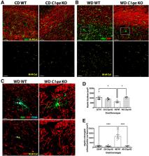
          Age-related cognitive decline and many dementias involve complex interactions of both genetic and environmental risk factors. Recent evidence has demonstrated a strong association of obesity with the development of dementia. Furthermore, white matter damage is found in obese subjects and mouse models of obesity. Here, we found that components of the complement cascade, including complement component 1qa (C1QA) and C3 are increased in the brain of Western diet (WD)-fed obese mice, particularly in white matter regions. To functionally test the role of the complement cascade in obesity-induced brain pathology, female and male mice deficient in C1QA, an essential molecule in the activation of the classical pathway of the complement cascade, were fed a WD and compared with WD-fed wild type (WT) mice, and to C1qa knock-out (KO) and WT mice fed a control diet (CD). C1qa KO mice fed a WD became obese but did not show pericyte loss or a decrease in laminin density in the cortex and hippocampus that was observed in obese WT controls. Furthermore, obesity-induced microglia phagocytosis and breakdown of myelin in the corpus callosum were also prevented by deficiency of C1QA. Collectively, these data show that C1QA is necessary for damage to the cerebrovasculature and white matter damage in diet-induced obesity.

          Related collections

          Most cited references30

          • Record: found
          • Abstract: found
          • Article: not found

          Will all Americans become overweight or obese? estimating the progression and cost of the US obesity epidemic.

          We projected future prevalence and BMI distribution based on national survey data (National Health and Nutrition Examination Study) collected between 1970s and 2004. Future obesity-related health-care costs for adults were estimated using projected prevalence, Census population projections, and published national estimates of per capita excess health-care costs of obesity/overweight. The objective was to illustrate potential burden of obesity prevalence and health-care costs of obesity and overweight in the United States that would occur if current trends continue. Overweight and obesity prevalence have increased steadily among all US population groups, but with notable differences between groups in annual increase rates. The increase (percentage points) in obesity and overweight in adults was faster than in children (0.77 vs. 0.46-0.49), and in women than in men (0.91 vs. 0.65). If these trends continue, by 2030, 86.3% adults will be overweight or obese; and 51.1%, obese. Black women (96.9%) and Mexican-American men (91.1%) would be the most affected. By 2048, all American adults would become overweight or obese, while black women will reach that state by 2034. In children, the prevalence of overweight (BMI >/= 95th percentile, 30%) will nearly double by 2030. Total health-care costs attributable to obesity/overweight would double every decade to 860.7-956.9 billion US dollars by 2030, accounting for 16-18% of total US health-care costs. We continue to move away from the Healthy People 2010 objectives. Timely, dramatic, and effective development and implementation of corrective programs/policies are needed to avoid the otherwise inevitable health and societal consequences implied by our projections .
            Bookmark
            • Record: found
            • Abstract: found
            • Article: found
            Is Open Access

            White matter hyperintensities in vascular contributions to cognitive impairment and dementia (VCID): Knowledge gaps and opportunities

            White matter hyperintensities (WMHs) are frequently seen on brain magnetic resonance imaging scans of older people. Usually interpreted clinically as a surrogate for cerebral small vessel disease, WMHs are associated with increased likelihood of cognitive impairment and dementia (including Alzheimer's disease [AD]). WMHs are also seen in cognitively healthy people. In this collaboration of academic, clinical, and pharmaceutical industry perspectives, we identify outstanding questions about WMHs and their relation to cognition, dementia, and AD. What molecular and cellular changes underlie WMHs? What are the neuropathological correlates of WMHs? To what extent are demyelination and inflammation present? Is it helpful to subdivide into periventricular and subcortical WMHs? What do WMHs signify in people diagnosed with AD? What are the risk factors for developing WMHs? What preventive and therapeutic strategies target WMHs? Answering these questions will improve prevention and treatment of WMHs and dementia.
              Bookmark
              • Record: found
              • Abstract: found
              • Article: found
              Is Open Access

              Cell-specific deletion of C1qa identifies microglia as the dominant source of C1q in mouse brain

              Background The complement cascade not only provides protection from infection but can also mediate destructive inflammation. Complement is also involved in elimination of neuronal synapses which is essential for proper development, but can be detrimental during aging and disease. C1q, required for several of these complement-mediated activities, is present in the neuropil, microglia, and a subset of interneurons in the brain. Methods To identify the source(s) of C1q in the brain, the C1qa gene was selectively inactivated in the microglia or Thy-1+ neurons in both wild type mice and a mouse model of Alzheimer’s disease (AD), and C1q synthesis assessed by immunohistochemistry, QPCR, and western blot analysis. Results While C1q expression in the brain was unaffected after inactivation of C1qa in Thy-1+ neurons, the brains of C1qa FL/FL :Cx3cr1 CreERT2 mice in which C1qa was ablated in microglia were devoid of C1q with the exception of limited C1q in subsets of interneurons. Surprisingly, this loss of C1q occurred even in the absence of tamoxifen by 1 month of age, demonstrating that Cre activity is tamoxifen-independent in microglia in Cx3cr1 CreERT2/WganJ mice. C1q expression in C1qa FL/FL : Cx3cr1 CreERT2/WganJ mice continued to decline and remained almost completely absent through aging and in AD model mice. No difference in C1q was detected in the liver or kidney from C1qa FL/FL : Cx3cr1 CreERT2/WganJ mice relative to controls, and C1qa FL/FL : Cx3cr1 CreERT2/WganJ mice had minimal, if any, reduction in plasma C1q. Conclusions Thus, microglia, but not neurons or peripheral sources, are the dominant source of C1q in the brain. While demonstrating that the Cx3cr1 CreERT2/WganJ deleter cannot be used for adult-induced deletion of genes in microglia, the model described here enables further investigation of physiological roles of C1q in the brain and identification of therapeutic targets for the selective control of complement-mediated activities contributing to neurodegenerative disorders. Electronic supplementary material The online version of this article (doi:10.1186/s12974-017-0814-9) contains supplementary material, which is available to authorized users.
                Bookmark

                Author and article information

                Journal
                eNeuro
                eNeuro
                eneuro
                eneuro
                eNeuro
                eNeuro
                Society for Neuroscience
                2373-2822
                9 April 2020
                2 June 2020
                May-Jun 2020
                : 7
                : 3
                : ENEURO.0057-20.2020
                Affiliations
                [1 ]The Jackson Laboratory , Bar Harbor, ME 04609
                [2 ]Graduate School of Biomedical Sciences, Tufts University School of Medicine , Boston, MA 02111
                [3 ]Department of Molecular & Cellular Biology, Rowan University , Glassboro, NJ 08028
                [4 ]Graduate School of Biomedical Sciences and Engineering, University of Maine , Orono, MA 04469
                Author notes

                The authors declare no competing financial interests.

                Author contributions: L.C.G. and G.R.H. designed research; L.C.G., H.E.K., I.S., and G.R.H. performed research; L.C.G., H.E.K., I.S., and G.R.H. analyzed data; L.C.G., H.E.K., I.S., and G.R.H. wrote the paper.

                This work was supported by the National Institutes on Aging Grant RF1 AG051496 (to G.R.H.).

                Correspondence should be addressed to Gareth R. Howell at gareth.howell@ 123456jax.org .
                Author information
                https://orcid.org/0000-0002-8380-2708
                Article
                eN-CFN-0057-20
                10.1523/ENEURO.0057-20.2020
                7294467
                32273396
                41899caf-d9ac-4f66-b36f-90c3682afdaa
                Copyright © 2020 Graham et al.

                This is an open-access article distributed under the terms of the Creative Commons Attribution 4.0 International license, which permits unrestricted use, distribution and reproduction in any medium provided that the original work is properly attributed.

                History
                : 17 February 2020
                : 22 February 2020
                Page count
                Figures: 8, Tables: 0, Equations: 0, References: 43, Pages: 12, Words: 00
                Funding
                Funded by: http://doi.org/10.13039/100000049HHS | NIH | National Institute on Aging (NIA)
                Categories
                3
                Research Article: Confirmation
                Disorders of the Nervous System
                Custom metadata
                May/June 2020

                complement,microglia,mouse,myelin,obesity,vasculature
                complement, microglia, mouse, myelin, obesity, vasculature

                Comments

                Comment on this article