21
views
0
recommends
+1 Recommend
0 collections
    0
    shares
      • Record: found
      • Abstract: found
      • Article: found
      Is Open Access

      Complement C1q-induced activation of β-catenin signalling causes hypertensive arterial remodelling

      research-article

      Read this article at

      Bookmark
          There is no author summary for this article yet. Authors can add summaries to their articles on ScienceOpen to make them more accessible to a non-specialist audience.

          Abstract

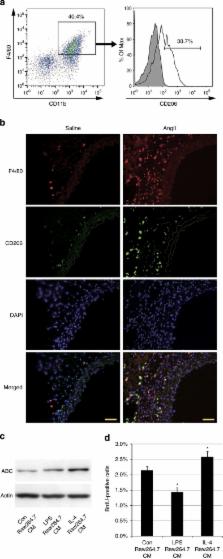
          Hypertension induces structural remodelling of arteries, which leads to arteriosclerosis and end-organ damage. Hyperplasia of vascular smooth muscle cells (VSMCs) and infiltration of immune cells are the hallmark of hypertensive arterial remodelling. However, the precise molecular mechanisms of arterial remodelling remain elusive. We have recently reported that complement C1q activates β-catenin signalling independent of Wnts. Here, we show a critical role of complement C1-induced activation of β-catenin signalling in hypertensive arterial remodelling. Activation of β-catenin and proliferation of VSMCs were observed after blood-pressure elevation, which were prevented by genetic and chemical inhibition of β-catenin signalling. Macrophage depletion and C1qa gene deletion attenuated the hypertension-induced β-catenin signalling, proliferation of VSMCs and pathological arterial remodelling. Our findings unveil the link between complement C1 and arterial remodelling and suggest that C1-induced activation of β-catenin signalling becomes a novel therapeutic target to prevent arteriosclerosis in patients with hypertension.

          Abstract

          The role of macrophages in hypertension-induced arterial remodeling is poorly understood. Here, Sumida et al. show that high blood pressure drives the alternatively activated macrophages to secrete complement C1q protein, which in turn elicits proliferative β-catenin signalling in the arterial smooth muscle cells.

          Related collections

          Most cited references24

          • Record: found
          • Abstract: found
          • Article: not found

          Proximal events in Wnt signal transduction.

          The Wnt family of secreted ligands act through many receptors to stimulate distinct intracellular signalling pathways in embryonic development, in adults and in disease processes. Binding of Wnt to the Frizzled family of receptors and to low density lipoprotein receptor-related protein 5 (LRP5) or LRP6 co-receptors stimulates the intracellular Wnt-beta-catenin signalling pathway, which regulates beta-cateninstability and context-dependent transcription. This signalling pathway controls many processes, such as cell fate determination, cell proliferation and self-renewal of stem and progenitor cells. Intriguingly, the transmembrane receptor Tyr kinases Ror2 and Ryk, as well as Frizzledreceptors that act independently of LRP5 or LRP6, function as receptors for Wnt and activate beta-catenin-independent pathways. This leads to changes in cell movement and polarity and to the antagonism of the beta-catenin pathway.
            Bookmark
            • Record: found
            • Abstract: found
            • Article: not found

            Homozygous C1q deficiency causes glomerulonephritis associated with multiple apoptotic bodies.

            The complement system plays a paradoxical role in the development and expression of autoimmunity in humans. The activation of complement in systemic lupus erythematosus (SLE) contributes to tissue injury. In contrast, inherited deficiency of classical pathway components, particularly C1q (ref. 1), is powerfully associated with the development of SLE. This leads to the hypothesis that a physiological action of the early part of the classical pathway protects against the development of SLE (ref. 2) and implies that C1q may play a key role in this respect. C1q-deficient (C1qa-/-) mice were generated by gene targeting and monitored for eight months. C1qa-/- mice had increased mortality and higher titres of autoantibodies, compared with strain-matched controls. Of the C1qa-/- mice, 25% had glomerulonephritis with immune deposits and multiple apoptotic cell bodies. Among mice without glomerulonephritis, there were significantly greater numbers of glomerular apoptotic bodies in C1q-deficient mice compared with controls. The phenotype associated with C1q deficiency was modified by background genes. These findings are compatible with the hypothesis that C1q deficiency causes autoimmunity by impairment of the clearance of apoptotic cells.
              Bookmark
              • Record: found
              • Abstract: found
              • Article: not found

              An adventitial IL-6/MCP1 amplification loop accelerates macrophage-mediated vascular inflammation leading to aortic dissection in mice.

              Vascular inflammation contributes to cardiovascular diseases such as aortic aneurysm and dissection. However, the precise inflammatory pathways involved have not been clearly defined. We have shown here that subcutaneous infusion of Ang II, a vasopressor known to promote vascular inflammation, into older C57BL/6J mice induced aortic production of the proinflammatory cytokine IL-6 and the monocyte chemoattractant MCP-1. Production of these factors occurred predominantly in the tunica adventitia, along with macrophage recruitment, adventitial expansion, and development of thoracic and suprarenal aortic dissections. In contrast, a reduced incidence of dissections was observed after Ang II infusion into mice lacking either IL-6 or the MCP-1 receptor CCR2. Further analysis revealed that Ang II induced CCR2+CD14hiCD11bhiF4/80- macrophage accumulation selectively in aortic dissections and not in aortas from Il6-/- mice. Adoptive transfer of Ccr2+/+ monocytes into Ccr2-/- mice resulted in selective monocyte uptake into the ascending and suprarenal aorta in regions of enhanced ROS stress, with restoration of IL-6 secretion and increased incidence of dissection. In vitro, coculture of monocytes and aortic adventitial fibroblasts produced MCP-1- and IL-6-enriched conditioned medium that promoted differentiation of monocytes into macrophages, induced CD14 and CD11b upregulation, and induced MCP-1 and MMP-9 expression. These results suggest that leukocyte-fibroblast interactions in the aortic adventitia potentiate IL-6 production, inducing local monocyte recruitment and activation, thereby promoting MCP-1 secretion, vascular inflammation, ECM remodeling, and aortic destabilization.
                Bookmark

                Author and article information

                Journal
                Nat Commun
                Nat Commun
                Nature Communications
                Nature Pub. Group
                2041-1723
                26 February 2015
                : 6
                : 6241
                Affiliations
                [1 ]Department of Cardiovascular Medicine, The University of Tokyo Graduate School of Medicine , Tokyo 113-8655, Japan
                [2 ]CREST, Japan Science and Technology Agency , Tokyo 102-0075, Japan
                [3 ]Department of Cardiovascular Medicine, Osaka University Graduate School of Medicine , Osaka 565-0871, Japan
                [4 ]Department of Cardiovascular Biology and Medicine, Niigata University Graduate School of Medical and Dental Sciences , Niigata 951-8510, Japan
                [5 ]Department of Pharmacology, Max-Planck-Institute for Heart and Lung Research , Bad Nauheim D-61231, Germany
                [6 ]Department of Cell Biology, The Cancer Institute, Japanese Foundation for Cancer Research , Tokyo 135-8550, Japan
                [7 ]Centre for Complement and Inflammation Research, Department of Medicine, Imperial College London , London SW7 2AZ, UK
                [8 ]Department of Cardiovascular Medicine, Chiba University Graduate School of Medicine , Chiba 260-8670, Japan
                [9 ]Department of Medicine II, Kansai Medical University , Osaka 573-1191, Japan
                Author notes
                Article
                ncomms7241
                10.1038/ncomms7241
                4351572
                25716000
                43818f29-0c00-4c29-a82e-cd5c6cbc469c
                Copyright © 2015, Nature Publishing Group, a division of Macmillan Publishers Limited. All Rights Reserved.

                This work is licensed under a Creative Commons Attribution 4.0 International License. The images or other third party material in this article are included in the article’s Creative Commons license, unless indicated otherwise in the credit line; if the material is not included under the Creative Commons license, users will need to obtain permission from the license holder to reproduce the material. To view a copy of this license, visit http://creativecommons.org/licenses/by/4.0/

                History
                : 09 December 2013
                : 08 January 2015
                Categories
                Article

                Uncategorized
                Uncategorized

                Comments

                Comment on this article