11
views
0
recommends
+1 Recommend
0 collections
    0
    shares
      • Record: found
      • Abstract: found
      • Article: found
      Is Open Access

      Definitions and classification of malformations of cortical development: practical guidelines

      review-article

      Read this article at

      Bookmark
          There is no author summary for this article yet. Authors can add summaries to their articles on ScienceOpen to make them more accessible to a non-specialist audience.

          Abstract

          Based on consensus meetings as part of the European Network Neuro-MIG initiative, Severino et al. provide standardized terminology and classification for malformations of cortical development, as well as practical recommendations to aid radiologists and neurologists in interpreting brain MRI in these patients.

          Abstract

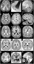
          Malformations of cortical development are a group of rare disorders commonly manifesting with developmental delay, cerebral palsy or seizures. The neurological outcome is extremely variable depending on the type, extent and severity of the malformation and the involved genetic pathways of brain development. Neuroimaging plays an essential role in the diagnosis of these malformations, but several issues regarding malformations of cortical development definitions and classification remain unclear. The purpose of this consensus statement is to provide standardized malformations of cortical development terminology and classification for neuroradiological pattern interpretation. A committee of international experts in paediatric neuroradiology prepared systematic literature reviews and formulated neuroimaging recommendations in collaboration with geneticists, paediatric neurologists and pathologists during consensus meetings in the context of the European Network Neuro-MIG initiative on Brain Malformations ( https://www.neuro-mig.org/). Malformations of cortical development neuroimaging features and practical recommendations are provided to aid both expert and non-expert radiologists and neurologists who may encounter patients with malformations of cortical development in their practice, with the aim of improving malformations of cortical development diagnosis and imaging interpretation worldwide.

          Related collections

          Most cited references136

          • Record: found
          • Abstract: found
          • Article: not found

          Measuring the thickness of the human cerebral cortex from magnetic resonance images.

          Accurate and automated methods for measuring the thickness of human cerebral cortex could provide powerful tools for diagnosing and studying a variety of neurodegenerative and psychiatric disorders. Manual methods for estimating cortical thickness from neuroimaging data are labor intensive, requiring several days of effort by a trained anatomist. Furthermore, the highly folded nature of the cortex is problematic for manual techniques, frequently resulting in measurement errors in regions in which the cortical surface is not perpendicular to any of the cardinal axes. As a consequence, it has been impractical to obtain accurate thickness estimates for the entire cortex in individual subjects, or group statistics for patient or control populations. Here, we present an automated method for accurately measuring the thickness of the cerebral cortex across the entire brain and for generating cross-subject statistics in a coordinate system based on cortical anatomy. The intersubject standard deviation of the thickness measures is shown to be less than 0.5 mm, implying the ability to detect focal atrophy in small populations or even individual subjects. The reliability and accuracy of this new method are assessed by within-subject test-retest studies, as well as by comparison of cross-subject regional thickness measures with published values.
            Bookmark
            • Record: found
            • Abstract: found
            • Article: not found

            Molecular identity of human outer radial glia during cortical development.

            Radial glia, the neural stem cells of the neocortex, are located in two niches: the ventricular zone and outer subventricular zone. Although outer subventricular zone radial glia may generate the majority of human cortical neurons, their molecular features remain elusive. By analyzing gene expression across single cells, we find that outer radial glia preferentially express genes related to extracellular matrix formation, migration, and stemness, including TNC, PTPRZ1, FAM107A, HOPX, and LIFR. Using dynamic imaging, immunostaining, and clonal analysis, we relate these molecular features to distinctive behaviors of outer radial glia, demonstrate the necessity of STAT3 signaling for their cell cycle progression, and establish their extensive proliferative potential. These results suggest that outer radial glia directly support the subventricular niche through local production of growth factors, potentiation of growth factor signals by extracellular matrix proteins, and activation of self-renewal pathways, thereby enabling the developmental and evolutionary expansion of the human neocortex.
              Bookmark
              • Record: found
              • Abstract: found
              • Article: found
              Is Open Access

              A developmental and genetic classification for malformations of cortical development: update 2012

              Malformations of cerebral cortical development include a wide range of developmental disorders that are common causes of neurodevelopmental delay and epilepsy. In addition, study of these disorders contributes greatly to the understanding of normal brain development and its perturbations. The rapid recent evolution of molecular biology, genetics and imaging has resulted in an explosive increase in our knowledge of cerebral cortex development and in the number and types of malformations of cortical development that have been reported. These advances continue to modify our perception of these malformations. This review addresses recent changes in our perception of these disorders and proposes a modified classification based upon updates in our knowledge of cerebral cortical development.
                Bookmark

                Author and article information

                Journal
                Brain
                Brain
                brainj
                Brain
                Oxford University Press
                0006-8950
                1460-2156
                October 2020
                10 August 2020
                10 August 2020
                : 143
                : 10
                : 2874-2894
                Affiliations
                [a1 ] Neuroradiology Unit, IRCCS Istituto Giannina Gaslini , Genoa, Italy
                [a2 ] Neuroradiology Unit, Imaging Department, Centro Hospitalar Vila Nova de Gaia/Espinho (CHVNG/E) , Vila Nova de Gaia, Portugal
                [a3 ] Department of Pediatric Radiology, HELIOS Klinikum Krefeld , Germany
                [a4 ] Division of Neuroradiology and Musculoskeletal Radiology, Department of Biomedical Imaging and Image-guided Therapy, Medical University of Vienna , Vienna, Austria
                [a5 ] Nalecz Institute of Biocybernetics and Biomedical Engineering Polish Academy of Sciences , Poland
                [a6 ] Neuroradiology Unit, Fondazione IRCCS Cà Granda, Ospedale Maggiore Policlinico, Department of Pathophysiology and Transplantation, Università degli Studi Milano , Italy
                [a7 ] Department of Neuroimaging Lab, Scientific Institute, IRCCS E. Medea , Bosisio Parini, Italy
                [a8 ] Department of Radiology, Great Ormond Street Hospital for Children NHS Foundation Trust , Great Ormond Street, London, UK
                [a9 ] Department of Neurology Royal Children’s Hospital, Murdoch Children’s Research Institute and University of Melbourne Department of Pediatrics , Melbourne, Australia
                [a10 ] Department of Clinical Genetics, Erasmus MC University Medical Center , Rotterdam, The Netherlands
                [a11 ] Department of Radiology and Biomedical Imaging, University of California , San Francisco, CA, USA
                [a12 ] Department of Neurology, University of California , San Francisco, CA, USA
                [a13 ] Department of Radiology, University Medical Center Utrecht , Utrecht, The Netherlands
                Author notes
                Correspondence to: Maarten H. Lequin Department of Radiology, Wilhelmina Children’s Hospital, University Medical Center Utrecht, Lundlaan 6, 3584 EA Utrecht, The Netherlands E-mail: M.H.Lequin@ 123456umcutrecht.nl

                James A. Barkovich, Maarten H. Lequin and Andrea Rossi contributed equally to this work.

                Author information
                http://orcid.org/0000-0003-4730-5322
                http://orcid.org/0000-0001-5465-3005
                http://orcid.org/0000-0002-1211-9979
                Article
                awaa174
                10.1093/brain/awaa174
                7586092
                32779696
                41744bb9-4adf-4e6d-b9ea-a4ce1c44e65c
                © The Author(s) (2020). Published by Oxford University Press on behalf of the Guarantors of Brain.

                This is an Open Access article distributed under the terms of the Creative Commons Attribution Non-Commercial License ( http://creativecommons.org/licenses/by-nc/4.0/), which permits non-commercial re-use, distribution, and reproduction in any medium, provided the original work is properly cited. For commercial re-use, please contact journals.permissions@oup.com

                History
                : 01 October 2019
                : 14 March 2020
                : 30 March 2020
                Page count
                Pages: 21
                Funding
                Funded by: European Cooperation in Science and Technology, DOI 10.13039/501100000921;
                Award ID: CA16118
                Categories
                Review Article
                AcademicSubjects/MED00310
                AcademicSubjects/SCI01870

                Neurosciences
                malformations of cortical development,classification,epilepsy,neuroimaging
                Neurosciences
                malformations of cortical development, classification, epilepsy, neuroimaging

                Comments

                Comment on this article