8
views
0
recommends
+1 Recommend
0 collections
    0
    shares
      • Record: found
      • Abstract: found
      • Article: found
      Is Open Access

      Tendon stem cell-derived exosomes regulate inflammation and promote the high-quality healing of injured tendon

      research-article

      Read this article at

      Bookmark
          There is no author summary for this article yet. Authors can add summaries to their articles on ScienceOpen to make them more accessible to a non-specialist audience.

          Abstract

          Background

          Tendon stem cells (TSCs) have been reported to hold promises for tendon repair and regeneration. However, less is known about the effects of exosomes derived from TSCs. Therefore, we aimed to clarify the healing effects of TSC-derived exosomes (TSC-Exos) on tendon injury.

          Methods

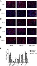
          The Achilles tendons of Sprague-Dawley male rats were used for primary culture of TSCs and tenocytes, and exosomes were isolated from TSCs. The proliferation of tenocytes induced by TSC-Exos was analyzed using an EdU assay; cell migration was measured by cell scratch and transwell assays. We used western blot to analyze the role of the PI3K/AKT and MAPK/ERK1/2 signaling pathways. In vivo, Achilles tendon injury models were created in Sprague-Dawley rats. Rats ( n = 54) were then randomly assigned to three groups: the TSC-Exos group, the GelMA group, and the control group. We used immunofluorescence to detect changes in the expression of inflammatory and apoptotic markers at 1 week after surgery. Histology and changes in expression of extracellular matrix (ECM)-related indices were assessed by hematoxylin-eosin (H&E) staining and immunohistochemistry at 2 and 8 weeks. The collagen fiber diameter of the healing tendon was analyzed at 8 weeks by transmission electron microscopy (TEM).

          Results

          TSC-Exos were taken up by tenocytes, which promoted the proliferation and migration of cells in a dose-dependent manner; this process may depend on the activation of the PI3K/AKT and MAPK/ERK1/2 signaling pathways. At 1 week after surgery, we found that inflammation and apoptosis were significantly suppressed by TSC-Exos. At 2 and 8 weeks, tendons treated with TSC-Exos showed more continuous and regular arrangement in contrast to disorganized tendons in the GelMA and control groups, and TSC-Exos may help regulate ECM balance and inhibited scar formation. Further, at 8 weeks, the TSC-Exos group had a larger diameter of collagen compared to the control group.

          Conclusions

          Our data suggest that TSC-Exos could promote high-quality healing of injured tendon, which may be a promising therapeutic approach for tendon injury.

          Related collections

          Most cited references42

          • Record: found
          • Abstract: found
          • Article: found
          Is Open Access

          Exosomes released from human induced pluripotent stem cells-derived MSCs facilitate cutaneous wound healing by promoting collagen synthesis and angiogenesis

          Background Human induced pluripotent stem cell-derived mesenchymal stem cells (hiPSC-MSCs) have emerged as a promising alternative for stem cell transplantation therapy. Exosomes derived from mesenchymal stem cells (MSC-Exos) can play important roles in repairing injured tissues. However, to date, no reports have demonstrated the use of hiPSC-MSC-Exos in cutaneous wound healing, and little is known regarding their underlying mechanisms in tissue repair. Methods hiPSC-MSC-Exos were injected subcutaneously around wound sites in a rat model and the efficacy of hiPSC-MSC-Exos was assessed by measuring wound closure areas, by histological and immunofluorescence examinations. We also evaluated the in vitro effects of hiPSC-MSC-Exos on both the proliferation and migration of human dermal fibroblasts and human umbilical vein endothelial cells (HUVECs) by cell-counting and scratch assays, respectively. The effects of exosomes on fibroblast collagen and elastin secretion were studied in enzyme-linked immunosorbent assays and quantitative reverse-transcriptase–polymerase chain reaction (qRT-PCR). In vitro capillary network formation was determined in tube-formation assays. Results Transplanting hiPSC-MSC-Exos to wound sites resulted in accelerated re-epithelialization, reduced scar widths, and the promotion of collagen maturity. Moreover, hiPSC-MSC-Exos not only promoted the generation of newly formed vessels, but also accelerated their maturation in wound sites. We found that hiPSC-MSC-Exos stimulated the proliferation and migration of human dermal fibroblasts and HUVECs in a dose-dependent manner in vitro. Similarly, Type I, III collagen and elastin secretion and mRNA expression by fibroblasts and tube formation by HUVECs were also increased with increasing hiPSC-MSC-Exos concentrations. Conclusions Our findings suggest that hiPSC-MSC-Exos can facilitate cutaneous wound healing by promoting collagen synthesis and angiogenesis. These data provide the first evidence for the potential of hiPSC-MSC-Exos in treating cutaneous wounds.
            Bookmark
            • Record: found
            • Abstract: found
            • Article: found
            Is Open Access

            Exosomes derived from human adipose mensenchymal stem cells accelerates cutaneous wound healing via optimizing the characteristics of fibroblasts

            Prolonged healing and scar formation are two major challenges in the treatment of soft tissue trauma. Adipose mesenchymal stem cells (ASCs) play an important role in tissue regeneration, and recent studies have suggested that exosomes secreted by stem cells may contribute to paracrine signaling. In this study, we investigated the roles of ASCs-derived exosomes (ASCs-Exos) in cutaneous wound healing. We found that ASCs-Exos could be taken up and internalized by fibroblasts to stimulate cell migration, proliferation and collagen synthesis in a dose-dependent manner, with increased genes expression of N-cadherin, cyclin-1, PCNA and collagen I, III. In vivo tracing experiments demonstrated that ASCs-Exos can be recruited to soft tissue wound area in a mouse skin incision model and significantly accelerated cutaneous wound healing. Histological analysis showed increased collagen I and III production by systemic administration of exosomes in the early stage of wound healing, while in the late stage, exosomes might inhibit collagen expression to reduce scar formation. Collectively, our findings indicate that ASCs-Exos can facilitate cutaneous wound healing via optimizing the characteristics of fibroblasts. Our results provide a new perspective and therapeutic strategy for the use of ASCs-Exos in soft tissue repair.
              Bookmark
              • Record: found
              • Abstract: found
              • Article: not found

              Biology of tendon injury: healing, modeling and remodeling.

              Tendon disorders are frequent, and are responsible for much morbidity both in sport and the workplace. Although the presence of degenerative changes does not always lead to symptoms, pre-existing degeneration has been implicated as a risk factor for acute tendon rupture. The term tendinopathy is a generic descriptor of the clinical conditions in and around tendons arising from overuse. The terms "tendinosis" and "tendinitis/tendonitis" should only be used after histopathological examination. Disordered healing is seen in tendinopathy, and inflammation is not typically seen. In acute injuries, the process of tendon healing is an indivisible process that can be categorized into three overlapping phases for descriptive purposes. Tendon healing can occur intrinsically, via proliferation of epitenon and endotenon tenocytes, or extrinsically, by invasion of cells from the surrounding sheath and synovium. Despite remodeling, the biochemical and mechanical properties of healed tendon tissue never match those of intact tendon. Tendon injuries account for considerable morbidity, and often prove disabling for several months, despite what is considered appropriate management. Chronic problems caused by overuse of tendons probably account for 30% of all running-related injuries, and the prevalence of elbow tendinopathy in tennis players can be as high as 40%. The basic cell biology of tendons is still not fully understood, and the management of tendon injury poses a considerable challenge for clinicians. This article describes the structure of tendons, and reviews the pathophysiology of tendon injury and healing.
                Bookmark

                Author and article information

                Contributors
                zhaozhu247@163.com
                Journal
                Stem Cell Res Ther
                Stem Cell Res Ther
                Stem Cell Research & Therapy
                BioMed Central (London )
                1757-6512
                17 September 2020
                17 September 2020
                2020
                : 11
                : 402
                Affiliations
                GRID grid.412463.6, ISNI 0000 0004 1762 6325, Department of Pediatric Surgery, , The Second Affiliated Hospital of Harbin Medical University, ; No. 246, Xuefu Road, Nangang District, Harbin, 150001 China
                Article
                1918
                10.1186/s13287-020-01918-x
                7499865
                32943109
                407d6862-fb4a-42ff-b134-d0d43577147a
                © The Author(s) 2020

                Open AccessThis article is licensed under a Creative Commons Attribution 4.0 International License, which permits use, sharing, adaptation, distribution and reproduction in any medium or format, as long as you give appropriate credit to the original author(s) and the source, provide a link to the Creative Commons licence, and indicate if changes were made. The images or other third party material in this article are included in the article's Creative Commons licence, unless indicated otherwise in a credit line to the material. If material is not included in the article's Creative Commons licence and your intended use is not permitted by statutory regulation or exceeds the permitted use, you will need to obtain permission directly from the copyright holder. To view a copy of this licence, visit http://creativecommons.org/licenses/by/4.0/. The Creative Commons Public Domain Dedication waiver ( http://creativecommons.org/publicdomain/zero/1.0/) applies to the data made available in this article, unless otherwise stated in a credit line to the data.

                History
                : 7 May 2020
                : 30 July 2020
                : 1 September 2020
                Funding
                Funded by: The National Natural Science Foundation of China and the Specialized Research Fund for Doctoral Programs in Colleges and Universities of China
                Award ID: 81871837, 81572117, 20132307110007
                Award Recipient :
                Categories
                Research
                Custom metadata
                © The Author(s) 2020

                Molecular medicine
                exosomes,tendon stem cells,proliferation,migration,inflammation,tendon healing
                Molecular medicine
                exosomes, tendon stem cells, proliferation, migration, inflammation, tendon healing

                Comments

                Comment on this article