4
views
0
recommends
+1 Recommend
0 collections
    0
    shares
      • Record: found
      • Abstract: found
      • Article: found
      Is Open Access

      Rescue of myocardial energetic dysfunction in diabetes through the correction of mitochondrial hyperacetylation by honokiol

      research-article

      Read this article at

      Bookmark
          There is no author summary for this article yet. Authors can add summaries to their articles on ScienceOpen to make them more accessible to a non-specialist audience.

          Abstract

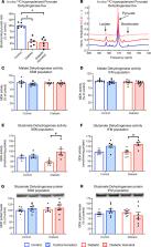
          Cardiac energetic dysfunction has been reported in patients with type 2 diabetes (T2D) and is an independent predictor of mortality. Identification of the mechanisms driving mitochondrial dysfunction, and therapeutic strategies to rescue these modifications, will improve myocardial energetics in T2D. We demonstrate using 31P-magnetic resonance spectroscopy ( 31P-MRS) that decreased cardiac ATP and phosphocreatine (PCr) concentrations occurred before contractile dysfunction or a reduction in PCr/ATP ratio in T2D. Real-time mitochondrial ATP synthesis rates and state 3 respiration rates were similarly depressed in T2D, implicating dysfunctional mitochondrial energy production. Driving this energetic dysfunction in T2D was an increase in mitochondrial protein acetylation, and increased ex vivo acetylation was shown to proportionally decrease mitochondrial respiration rates. Treating T2D rats in vivo with the mitochondrial deacetylase SIRT3 activator honokiol reversed the hyperacetylation of mitochondrial proteins and restored mitochondrial respiration rates to control levels. Using 13C-hyperpolarized MRS, respiration with different substrates, and enzyme assays, we localized this improvement to increased glutamate dehydrogenase activity. Finally, honokiol treatment increased ATP and PCr concentrations and increased total ATP synthesis flux in the T2D heart. In conclusion, hyperacetylation drives energetic dysfunction in T2D, and reversing acetylation with the SIRT3 activator honokiol rescued myocardial and mitochondrial energetics in T2D.

          Abstract

          Abstract

          Pharmacologically targeting mitochondrial acetylation provides a mechanism to rescue impaired myocardial energy generation in type 2 diabetes.

          Related collections

          Most cited references24

          • Record: found
          • Abstract: found
          • Article: not found

          SRT1720, SRT2183, SRT1460, and resveratrol are not direct activators of SIRT1.

          Sirtuins catalyze NAD(+)-dependent protein deacetylation and are critical regulators of transcription, apoptosis, metabolism, and aging. There are seven human sirtuins (SIRT1-7), and SIRT1 has been implicated as a key mediator of the pathways downstream of calorie restriction that have been shown to delay the onset and reduce the incidence of age-related diseases such as type 2 diabetes. Increasing SIRT1 activity, either by transgenic overexpression of the Sirt1 gene in mice or by pharmacological activation by small molecule activators resveratrol and SRT1720, has shown beneficial effects in rodent models of type 2 diabetes, indicating that SIRT1 may represent an attractive therapeutic target. Herein, we have assessed purported SIRT1 activators by employing biochemical assays utilizing native substrates, including a p53-derived peptide substrate lacking a fluorophore as well as the purified native full-length protein substrates p53 and acetyl-CoA synthetase1. SRT1720, its structurally related compounds SRT2183 and SRT1460, and resveratrol do not lead to apparent activation of SIRT1 with native peptide or full-length protein substrates, whereas they do activate SIRT1 with peptide substrate containing a covalently attached fluorophore. Employing NMR, surface plasmon resonance, and isothermal calorimetry techniques, we provide evidence that these compounds directly interact with fluorophore-containing peptide substrates. Furthermore, we demonstrate that SRT1720 neither lowers plasma glucose nor improves mitochondrial capacity in mice fed a high fat diet. SRT1720, SRT2183, SRT1460, and resveratrol exhibit multiple off-target activities against receptors, enzymes, transporters, and ion channels. Taken together, we conclude that SRT1720, SRT2183, SRT1460, and resveratrol are not direct activators of SIRT1.
            Bookmark
            • Record: found
            • Abstract: found
            • Article: not found

            Mitochondrial complex I deficiency increases protein acetylation and accelerates heart failure.

            Mitochondrial respiratory dysfunction is linked to the pathogenesis of multiple diseases, including heart failure, but the specific mechanisms for this link remain largely elusive. We modeled the impairment of mitochondrial respiration by the inactivation of the Ndufs4 gene, a protein critical for complex I assembly, in the mouse heart (cKO). Although complex I-supported respiration decreased by >40%, the cKO mice maintained normal cardiac function in vivo and high-energy phosphate content in isolated perfused hearts. However, the cKO mice developed accelerated heart failure after pressure overload or repeated pregnancy. Decreased NAD(+)/NADH ratio by complex I deficiency inhibited Sirt3 activity, leading to an increase in protein acetylation and sensitization of the permeability transition in mitochondria (mPTP). NAD(+) precursor supplementation to cKO mice partially normalized the NAD(+)/NADH ratio, protein acetylation, and mPTP sensitivity. These findings describe a mechanism connecting mitochondrial dysfunction to the susceptibility to diseases and propose a potential therapeutic target. Copyright © 2013 Elsevier Inc. All rights reserved.
              Bookmark
              • Record: found
              • Abstract: found
              • Article: not found

              Honokiol blocks and reverses cardiac hypertrophy in mice by activating mitochondrial SIRT3

              Honokiol (HKL) is a natural biphenolic compound derived from the bark of magnolia trees with anti-inflammatory, anti-oxidative, anti-tumor and neuroprotective properties. Here we show that HKL blocks agonist-induced and pressure overload-mediated, cardiac hypertrophic responses, and ameliorates pre-existing cardiac hypertrophy, in mice. Our data suggest that the anti-hypertrophic effects of HKL depend on activation of the deacetylase SIRT3. We demonstrate that HKL is present in mitochondria, enhances SIRT3 expression nearly two-fold and suggest that HKL may bind to SIRT3 to further increase its activity. Increased SIRT3 activity is associated with reduced acetylation of mitochondrial SIRT3 substrates, MnSOD and OSCP. HKL-treatment increases mitochondrial rate of oxygen consumption and reduces ROS synthesis in wild-type, but not in SIRT3-KO cells. Moreover, HKL-treatment blocks cardiac fibroblast proliferation and differentiation to myofibroblasts in SIRT3-dependent manner. These results suggest that HKL is a pharmacological activator of SIRT3 capable of blocking, and even reversing, the cardiac hypertrophic response.
                Bookmark

                Author and article information

                Contributors
                Journal
                JCI Insight
                JCI Insight
                JCI Insight
                JCI Insight
                American Society for Clinical Investigation
                2379-3708
                3 September 2020
                3 September 2020
                3 September 2020
                : 5
                : 17
                : e140326
                Affiliations
                [1 ]Department of Physiology, Anatomy and Genetics, University of Oxford, Oxford, United Kingdom.
                [2 ]Oxford Centre for Clinical Magnetic Resonance Research, John Radcliffe Hospital, Oxford, United Kingdom.
                [3 ]Department of Physics, University of Oxford, Oxford, United Kingdom.
                [4 ]Vascular Medicine Institute, University of Pittsburgh, Pittsburgh, Pennsylvania, USA.
                Author notes
                Address correspondence to: Lisa Heather, Department of Physiology, Anatomy and Genetics, Sherrington Building, University of Oxford, Parks Road, Oxford, OX1 3PT, United Kingdom. Phone: 44.1865.282048; Email: lisa.heather@ 123456dpag.ox.ac.uk .
                Author information
                http://orcid.org/0000-0002-6258-1299
                Article
                140326
                10.1172/jci.insight.140326
                7526448
                32879143
                3edbe91b-0154-4404-b1e0-d65eb34e3dbe
                © 2020 Kerr et al.

                This work is licensed under the Creative Commons Attribution 4.0 International License. To view a copy of this license, visit http://creativecommons.org/licenses/by/4.0/.

                History
                : 15 May 2020
                : 16 July 2020
                Funding
                Funded by: British Heart Foundation, https://doi.org/10.13039/501100000274;
                Award ID: FS/17/58/33072
                Categories
                Research Article

                cardiology,metabolism,mitochondria
                cardiology, metabolism, mitochondria

                Comments

                Comment on this article