0
views
0
recommends
+1 Recommend
0 collections
    0
    shares
      • Record: found
      • Abstract: found
      • Article: found
      Is Open Access

      Real-world effects of anti-vascular endothelial growth factor injection frequency on visual outcomes in patients with diabetic macular oedema

      research-article

      Read this article at

      Bookmark
          There is no author summary for this article yet. Authors can add summaries to their articles on ScienceOpen to make them more accessible to a non-specialist audience.

          Abstract

          Background and objective

          Anti-vascular endothelial growth factor (VEGF) injections are often administered less frequently in real-world treatment of diabetic macular oedema (DMO) than what was studied in clinical trials. This study aims to characterise real-world DMO treatment patterns and the effect of treatment intervals on patient outcomes.

          Study design/patients and methods

          This was a retrospective study of 291 patients with DMO treated with anti-VEGF therapy. 12- and 24-month best visual acuity (BVA) and central subfield thickness (CST) were compared between injection interval groups, which were determined by averaging the two most recent injection intervals. Multiple linear regressions were performed to identify factors associated with injection interval, BVA, and CST.

          Results

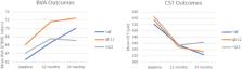
          48.8% of patients received injections less than or equal to every 8 weeks (≤ q8w), 27.5% between every 8 to 12 weeks (q8–12w), and 23.7% greater than every 12 weeks (> q12w). Baseline CST was similar ( p = 0.32), but BVA differed significantly in q8–12w patients ( p = 0.0095). BVA and CST at 12 months were similar, but q8–12w patients experienced greater 12-month BVA improvement (7.36 ± 12.4 letters) than > q12w patients (1.26 ± 12.3 letters; p = 0.0056). 24-month BVA and CST changes were similar between groups ( p = 0.30 and 0.87). Baseline BVA, HbA1c, and sex were associated with 12-month BVA, and baseline BVA and CST were associated with 12-month CST.

          Conclusion

          Many patients experienced improvements in BVA and CST over 12 months of treatment despite receiving less frequent anti-VEGF therapy than recommended in the pivotal trials. The present study showed that extended treatment intervals with bevacizumab were effective in preserving vision of many individuals with high baseline BVA.

          Related collections

          Most cited references21

          • Record: found
          • Abstract: found
          • Article: found
          Is Open Access

          Global Prevalence and Major Risk Factors of Diabetic Retinopathy

          OBJECTIVE To examine the global prevalence and major risk factors for diabetic retinopathy (DR) and vision-threatening diabetic retinopathy (VTDR) among people with diabetes. RESEARCH DESIGN AND METHODS A pooled analysis using individual participant data from population-based studies around the world was performed. A systematic literature review was conducted to identify all population-based studies in general populations or individuals with diabetes who had ascertained DR from retinal photographs. Studies provided data for DR end points, including any DR, proliferative DR, diabetic macular edema, and VTDR, and also major systemic risk factors. Pooled prevalence estimates were directly age-standardized to the 2010 World Diabetes Population aged 20–79 years. RESULTS A total of 35 studies (1980–2008) provided data from 22,896 individuals with diabetes. The overall prevalence was 34.6% (95% CI 34.5–34.8) for any DR, 6.96% (6.87–7.04) for proliferative DR, 6.81% (6.74–6.89) for diabetic macular edema, and 10.2% (10.1–10.3) for VTDR. All DR prevalence end points increased with diabetes duration, hemoglobin A1c, and blood pressure levels and were higher in people with type 1 compared with type 2 diabetes. CONCLUSIONS There are approximately 93 million people with DR, 17 million with proliferative DR, 21 million with diabetic macular edema, and 28 million with VTDR worldwide. Longer diabetes duration and poorer glycemic and blood pressure control are strongly associated with DR. These data highlight the substantial worldwide public health burden of DR and the importance of modifiable risk factors in its occurrence. This study is limited by data pooled from studies at different time points, with different methodologies and population characteristics.
            Bookmark
            • Record: found
            • Abstract: found
            • Article: not found

            Ranibizumab for diabetic macular edema: results from 2 phase III randomized trials: RISE and RIDE.

            To evaluate the efficacy and safety of intravitreal ranibizumab in diabetic macular edema (DME) patients. Two parallel, methodologically identical, phase III, multicenter, double-masked, sham injection-controlled, randomized studies. Adults with vision loss from DME (best-corrected visual acuity [BCVA], 20/40-20/320 Snellen equivalent) and central subfield thickness ≥275 μm on time-domain optical coherence tomography (OCT). Monthly intravitreal ranibizumab (0.5 or 0.3 mg) or sham injections. Macular laser was available per-protocol-specified criteria. Proportion of patients gaining ≥15 letters in BCVA from baseline at 24 months. In RISE (NCT00473330), 377 patients were randomized (127 to sham, 125 to 0.3 mg, 125 to 0.5 mg). At 24 months, 18.1% of sham patients gained ≥15 letters versus 44.8% of 0.3-mg (P<0.0001; difference vs sham adjusted for randomization stratification factors, 24.3%; 95% confidence interval [CI], 13.8-34.8) and 39.2% of 0.5-mg ranibizumab patients (P<0.001; adjusted difference, 20.9%; 95% CI, 10.7-31.1). In RIDE (NCT00473382), 382 patients were randomized (130 to sham, 125 to 0.3 mg, 127 to 0.5 mg). Significantly more ranibizumab-treated patients gained ≥15 letters: 12.3% of sham patients versus 33.6% of 0.3-mg patients (P<0.0001; adjusted difference, 20.8%; 95% CI, 11.4-30.2) and 45.7% of 0.5-mg ranibizumab patients (P<0.0001; adjusted difference, 33.3%; 95% CI, 23.8-42.8). Significant improvements in macular edema were noted on OCT, and retinopathy was less likely to worsen and more likely to improve in ranibizumab-treated patients. Ranibizumab-treated patients underwent significantly fewer macular laser procedures (mean of 1.8 and 1.6 laser procedures over 24 months in the sham groups vs 0.3-0.8 in ranibizumab groups). Ocular safety was consistent with prior ranibizumab studies; endophthalmitis occurred in 4 ranibizumab patients. The total incidence of deaths from vascular or unknown causes, nonfatal myocardial infarctions, and nonfatal cerebrovascular accidents, which are possible effects from systemic vascular endothelial growth factor inhibition, was 4.9% to 5.5% of sham patients and 2.4% to 8.8% of ranibizumab patients. Ranibizumab rapidly and sustainably improved vision, reduced the risk of further vision loss, and improved macular edema in patients with DME, with low rates of ocular and nonocular harm. Copyright © 2012 American Academy of Ophthalmology. Published by Elsevier Inc. All rights reserved.
              Bookmark
              • Record: found
              • Abstract: found
              • Article: not found

              Prevalence of and risk factors for diabetic macular edema in the United States.

              Diabetic macular edema (DME) is a leading cause of vision loss in persons with diabetes mellitus. Although there are national estimates for the prevalence of diabetic retinopathy and its risk factors among persons with diabetes, to our knowledge, no comparable estimates are available for DME specifically.
                Bookmark

                Author and article information

                Contributors
                talcotk@ccf.org
                Journal
                Eye (Lond)
                Eye (Lond)
                Eye
                Nature Publishing Group UK (London )
                0950-222X
                1476-5454
                6 March 2024
                6 March 2024
                June 2024
                : 38
                : 9
                : 1687-1693
                Affiliations
                [1 ]Case Western Reserve University School of Medicine, ( https://ror.org/051fd9666) Cleveland, OH USA
                [2 ]Cleveland Clinic Cole Eye Institute Center for Ophthalmic Bioinformatics, ( https://ror.org/03xjacd83) Cleveland, OH USA
                [3 ]Cleveland Clinic Lerner College of Medicine, ( https://ror.org/02x4b0932) Cleveland, OH USA
                Author information
                http://orcid.org/0000-0001-9227-9560
                http://orcid.org/0000-0001-6197-9476
                http://orcid.org/0000-0001-5859-8162
                Article
                2998
                10.1038/s41433-024-02998-2
                11156885
                38448732
                3e6fdf75-d0f2-40d1-8957-8dc571515ea8
                © The Author(s) 2024

                Open Access This article is licensed under a Creative Commons Attribution 4.0 International License, which permits use, sharing, adaptation, distribution and reproduction in any medium or format, as long as you give appropriate credit to the original author(s) and the source, provide a link to the Creative Commons licence, and indicate if changes were made. The images or other third party material in this article are included in the article’s Creative Commons licence, unless indicated otherwise in a credit line to the material. If material is not included in the article’s Creative Commons licence and your intended use is not permitted by statutory regulation or exceeds the permitted use, you will need to obtain permission directly from the copyright holder. To view a copy of this licence, visit http://creativecommons.org/licenses/by/4.0/.

                History
                : 13 October 2022
                : 16 January 2024
                : 15 February 2024
                Categories
                Article
                Custom metadata
                © The Royal College of Ophthalmologists 2024

                Vision sciences
                outcomes research,retinal diseases
                Vision sciences
                outcomes research, retinal diseases

                Comments

                Comment on this article