10
views
0
recommends
+1 Recommend
0 collections
    0
    shares
      • Record: found
      • Abstract: found
      • Article: found
      Is Open Access

      Rho-Kinase Inhibitors for the Treatment of Refractory Diabetic Macular Oedema

      review-article

      Read this article at

      Bookmark
          There is no author summary for this article yet. Authors can add summaries to their articles on ScienceOpen to make them more accessible to a non-specialist audience.

          Abstract

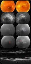
          Diabetic macular oedema (DMO) is one of the leading causes of vision loss associated with diabetic retinopathy (DR). New insights in managing this condition have changed the paradigm in its treatment, with intravitreal injections of antivascular endothelial growth factor (anti-VEGF) having become the standard therapy for DMO worldwide. However, there is no single standard therapy for all patients DMO refractory to anti-VEGF treatment; thus, further investigation is still needed. The key obstacles in developing suitable therapeutics for refractory DMO lie in its complex pathophysiology; therefore, there is an opportunity for further improvements in the progress and applications of new drugs. Previous studies have indicated that Rho-associated kinase (Rho-kinase/ROCK) is an essential molecule in the pathogenesis of DMO. This is why the Rho/ROCK signalling pathway has been proposed as a possible target for new treatments. The present review focuses on the recent progress on the possible role of ROCK and its therapeutic potential in DMO. A systematic literature search was performed, covering the years 1991 to 2021, using the following keywords: “rho-Associated Kinas-es”, “Diabetic Retinopathy”, “Macular Edema”, “Ripasudil”, “Fasudil” and “Netarsudil”. Better insight into the pathological role of Rho-kinase/ROCK may lead to the development of new strategies for refractory DMO treatment and prevention.

          Related collections

          Most cited references149

          • Record: found
          • Abstract: found
          • Article: found
          Is Open Access

          Global Prevalence and Major Risk Factors of Diabetic Retinopathy

          OBJECTIVE To examine the global prevalence and major risk factors for diabetic retinopathy (DR) and vision-threatening diabetic retinopathy (VTDR) among people with diabetes. RESEARCH DESIGN AND METHODS A pooled analysis using individual participant data from population-based studies around the world was performed. A systematic literature review was conducted to identify all population-based studies in general populations or individuals with diabetes who had ascertained DR from retinal photographs. Studies provided data for DR end points, including any DR, proliferative DR, diabetic macular edema, and VTDR, and also major systemic risk factors. Pooled prevalence estimates were directly age-standardized to the 2010 World Diabetes Population aged 20–79 years. RESULTS A total of 35 studies (1980–2008) provided data from 22,896 individuals with diabetes. The overall prevalence was 34.6% (95% CI 34.5–34.8) for any DR, 6.96% (6.87–7.04) for proliferative DR, 6.81% (6.74–6.89) for diabetic macular edema, and 10.2% (10.1–10.3) for VTDR. All DR prevalence end points increased with diabetes duration, hemoglobin A1c, and blood pressure levels and were higher in people with type 1 compared with type 2 diabetes. CONCLUSIONS There are approximately 93 million people with DR, 17 million with proliferative DR, 21 million with diabetic macular edema, and 28 million with VTDR worldwide. Longer diabetes duration and poorer glycemic and blood pressure control are strongly associated with DR. These data highlight the substantial worldwide public health burden of DR and the importance of modifiable risk factors in its occurrence. This study is limited by data pooled from studies at different time points, with different methodologies and population characteristics.
            Bookmark
            • Record: found
            • Abstract: found
            • Article: not found

            A central role for inflammation in the pathogenesis of diabetic retinopathy.

            Diabetic retinopathy is a leading cause of adult vision loss and blindness. Much of the retinal damage that characterizes the disease results from retinal vascular leakage and nonperfusion. Diabetic retinal vascular leakage, capillary nonperfusion, and endothelial cell damage are temporary and spatially associated with retinal leukocyte stasis in early experimental diabetes. Retinal leukostasis increases within days of developing diabetes and correlates with the increased expression of retinal intercellular adhesion molecule-1 (ICAM-1) and CD18. Mice deficient in the genes encoding for the leukocyte adhesion molecules CD18 and ICAM-1 were studied in two models of diabetic retinopathy with respect to the long-term development of retinal vascular lesions. CD18-/- and ICAM-1-/- mice demonstrate significantly fewer adherent leukocytes in the retinal vasculature at 11 and 15 months after induction of diabetes with STZ. This condition is associated with fewer damaged endothelial cells and lesser vascular leakage. Galactosemia of up to 24 months causes pericyte and endothelial cell loss and formation of acellular capillaries. These changes are significantly reduced in CD18- and ICAM-1-deficient mice. Basement membrane thickening of the retinal vessels is increased in long-term galactosemic animals independent of the genetic strain. Here we show that chronic, low-grade subclinical inflammation is responsible for many of the signature vascular lesions of diabetic retinopathy. These data highlight the central and causal role of adherent leukocytes in the pathogenesis of diabetic retinopathy. They also underscore the potential utility of anti-inflammatory treatment in diabetic retinopathy.
              Bookmark
              • Record: found
              • Abstract: found
              • Article: not found

              Aflibercept, bevacizumab, or ranibizumab for diabetic macular edema.

              The relative efficacy and safety of intravitreous aflibercept, bevacizumab, and ranibizumab in the treatment of diabetic macular edema are unknown.
                Bookmark

                Author and article information

                Contributors
                Role: Academic Editor
                Journal
                Cells
                Cells
                cells
                Cells
                MDPI
                2073-4409
                03 July 2021
                July 2021
                : 10
                : 7
                : 1683
                Affiliations
                [1 ]Department of Ophthalmology, Hospital Clínico Universitario de Valladolid, 47003 Valladolid, Spain; mmateoso@ 123456saludcastillayleon.es (M.M.-O.); Luis.GarciaOnrubia@ 123456gstt.nhs.uk (L.G.-O.); fvalentin@ 123456saludcastillayleon.es (F.J.V.-B.); mlopezgal@ 123456saludcastillayleon.es (M.L.-G.); pastor@ 123456ioba.med.uva.es (J.C.P.)
                [2 ]Department of Ophthalmology, St Thomas’ Hospital, London SE1 7EH, UK
                [3 ]Area of Infectious, Inflammatory and Metabolic Disease, Institute of Biomedical Research of Salamanca (IBSAL), 37007 Salamanca, Spain; gonzalez@ 123456usal.es
                [4 ]Institute of Molecular and Cellular Biology of Cancer (IBMCC), University of Salamanca-CSIC, 37007 Salamanca, Spain
                [5 ]Retina Group, IOBA (Institute of Applied Ophthalmobiology), University of Valladolid, 47002 Valladolid, Spain
                [6 ]Cooperative Network for Research in Ophthalmology Oftared, National Institute of Health Carlos III, 28220 Madrid, Spain
                Author notes
                [* ]Correspondence: rusateguim@ 123456ioba.med.uva.es (R.U.-M.); spastori@ 123456ioba.med.uva.es (S.P.-I.); Tel.: +34-983-423-559
                Author information
                https://orcid.org/0000-0002-2726-6795
                https://orcid.org/0000-0001-5934-7306
                Article
                cells-10-01683
                10.3390/cells10071683
                8307715
                1089e3cc-54b7-4207-ad33-c8f62b0bfe54
                © 2021 by the authors.

                Licensee MDPI, Basel, Switzerland. This article is an open access article distributed under the terms and conditions of the Creative Commons Attribution (CC BY) license ( https://creativecommons.org/licenses/by/4.0/).

                History
                : 17 May 2021
                : 29 June 2021
                Categories
                Review

                diabetic macular oedema,diabetic retinopathy,macular oedema,rho-associated kinases

                Comments

                Comment on this article