61
views
0
recommends
+1 Recommend
0 collections
    0
    shares
      • Record: found
      • Abstract: found
      • Article: found
      Is Open Access

      Ultrafast spontaneous emission source using plasmonic nanoantennas

      research-article

      Read this article at

      Bookmark
          There is no author summary for this article yet. Authors can add summaries to their articles on ScienceOpen to make them more accessible to a non-specialist audience.

          Abstract

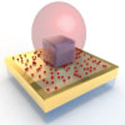
          Typical emitters such as molecules, quantum dots and semiconductor quantum wells have slow spontaneous emission with lifetimes of 1–10 ns, creating a mismatch with high-speed nanoscale optoelectronic devices such as light-emitting diodes, single-photon sources and lasers. Here we experimentally demonstrate an ultrafast (<11 ps) yet efficient source of spontaneous emission, corresponding to an emission rate exceeding 90 GHz, using a hybrid structure of single plasmonic nanopatch antennas coupled to colloidal quantum dots. The antennas consist of silver nanocubes coupled to a gold film separated by a thin polymer spacer layer and colloidal core–shell quantum dots, a stable and technologically relevant emitter. We show an increase in the spontaneous emission rate of a factor of 880 and simultaneously a 2,300-fold enhancement in the total fluorescence intensity, which indicates a high radiative quantum efficiency of ∼50%. The nanopatch antenna geometry can be tuned from the visible to the near infrared, providing a promising approach for nanophotonics based on ultrafast spontaneous emission.

          Abstract

          Typical emitters such as molecules and quantum dots have slow spontaneous emission with lifetimes of 1–10 ns. Here, Hoang et al. have fabricated a hybrid structure of plasmonic nanopatch antennas coupled to quantum dots, achieving ultrafast spontaneous emission with a lifetime of 11 ps.

          Related collections

          Most cited references27

          • Record: found
          • Abstract: found
          • Article: found
          Is Open Access

          Quantum nature of a strongly-coupled single quantum dot-cavity system

          , , (2006)
          Cavity quantum electrodynamics (QED) studies the interaction between a quantum emitter and a single radiation-field mode. When an atom is in strong coupling with a cavity mode1,2, it is possible to realize key quantum information processing (QIP) tasks, such as controlled coherent coupling and entanglement of distinguishable quantum systems. Realizing these tasks in the solid state is clearly desirable, and coupling semiconductor self-assembled quantum dots (QDs) to monolithic optical cavities is a promising route to this end. However, validating the efficacy of QDs in QIP applications requires confirmation of the quantum nature of the QD-cavity system in the strong coupling regime. Here we find a confirmation by observing quantum correlations in photoluminescence (PL) from a photonic crystal (PC) nanocavity3-5 interacting with one, and only one, QD located precisely at the cavity electric field maximum. When off-resonance, photon emission from the cavity mode and QD excitons is anti-correlated at the level of single quanta, proving that the mode is driven solely by the QD despite an energy mis-match between cavity and excitons. When tuned into resonance, the exciton and photon enter the strong-coupling regime of cavity-QED and the QD lifetime reduces by a factor of 120. The photon stream from the cavity becomes anti-bunched, proving that the coupled exciton/photon system is in the quantum anharmonic regime. Our observations unequivocally show that QIP tasks requiring the quantum nonlinear regime are achievable in the solid state.
            Bookmark
            • Record: found
            • Abstract: found
            • Article: not found

            Lasing action in strongly coupled plasmonic nanocavity arrays.

            Periodic dielectric structures are typically integrated with a planar waveguide to create photonic band-edge modes for feedback in one-dimensional distributed feedback lasers and two-dimensional photonic-crystal lasers. Although photonic band-edge lasers are widely used in optics and biological applications, drawbacks include low modulation speeds and diffraction-limited mode confinement. In contrast, plasmonic nanolasers can support ultrafast dynamics and ultrasmall mode volumes. However, because of the large momentum mismatch between their nanolocalized lasing fields and free-space light, they suffer from large radiative losses and lack beam directionality. Here, we report lasing action from band-edge lattice plasmons in arrays of plasmonic nanocavities in a homogeneous dielectric environment. We find that optically pumped, two-dimensional arrays of plasmonic Au or Ag nanoparticles surrounded by an organic gain medium show directional beam emission (divergence angle <1.5° and linewidth <1.3 nm) characteristic of lasing action in the far-field, and behave as arrays of nanoscale light sources in the near-field. Using a semi-quantum electromagnetic approach to simulate the active optical responses, we show that lasing is achieved through stimulated energy transfer from the gain to the band-edge lattice plasmons in the deep subwavelength vicinity of the individual nanoparticles. Using femtosecond-transient absorption spectroscopy, we verified that lattice plasmons in plasmonic nanoparticle arrays could reach a 200-fold enhancement of the spontaneous emission rate of the dye because of their large local density of optical states.
              Bookmark
              • Record: found
              • Abstract: found
              • Article: not found

              Plasmonic enhancement of molecular fluorescence.

              Metallic nanoparticles are known to dramatically modify the spontaneous emission of nearby fluorescent molecules and materials. Here we examine the role of the nanoparticle plasmon resonance energy and nanoparticle scattering cross section on the fluorescence enhancement of adjacent indocyanine green (ICG) dye molecules. We find that enhancement of the molecular fluorescence by more than a factor of 50 can be achieved for ICG next to a nanoparticle with a large scattering cross section and a plasmon resonance frequency corresponding to the emission frequency of the molecule.
                Bookmark

                Author and article information

                Journal
                Nat Commun
                Nat Commun
                Nature Communications
                Nature Pub. Group
                2041-1723
                27 July 2015
                2015
                : 6
                : 7788
                Affiliations
                [1 ]Department of Physics, Duke University , Durham, North Carolina 27708, USA.
                [2 ]Center for Metamaterials and Integrated Plasmonics, Duke University , Durham, North Carolina 27708, USA.
                [3 ]Department of Electrical and Computer Engineering, Duke University , Durham, North Carolina 27708, USA.
                Author notes
                [*]

                These authors contributed equally to this work.

                [†]

                Present address: Department of Electrical and Computer Engineering, University of Nebraska-Lincoln, Lincoln, Nebraska 68588-0511, USA.

                Author information
                http://orcid.org/0000-0002-0487-7585
                Article
                ncomms8788
                10.1038/ncomms8788
                4525280
                26212857
                3be74694-f580-4b8d-b04a-55c32f3dcc1d
                Copyright © 2015, Nature Publishing Group, a division of Macmillan Publishers Limited. All Rights Reserved.

                This work is licensed under a Creative Commons Attribution 4.0 International License. The images or other third party material in this article are included in the article's Creative Commons license, unless indicated otherwise in the credit line; if the material is not included under the Creative Commons license, users will need to obtain permission from the license holder to reproduce the material. To view a copy of this license, visit http://creativecommons.org/licenses/by/4.0/

                History
                : 01 March 2015
                : 11 June 2015
                Categories
                Article

                Uncategorized
                Uncategorized

                Comments

                Comment on this article