14
views
0
recommends
+1 Recommend
0 collections
    0
    shares
      • Record: found
      • Abstract: found
      • Article: found
      Is Open Access

      Absence Seizures as a Feature of Juvenile Myoclonic Epilepsy in Rhodesian Ridgeback Dogs

      case-report

      Read this article at

      Bookmark
          There is no author summary for this article yet. Authors can add summaries to their articles on ScienceOpen to make them more accessible to a non-specialist audience.

          Abstract

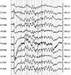
          Myoclonic epilepsy in Rhodesian Ridgeback (RR) dogs is characterized by myoclonic seizures occurring mainly during relaxation periods, a juvenile age of onset and generalized tonic‐clonic seizures in one‐third of patients. An 8‐month‐old female intact RR was presented for myoclonic seizures and staring episodes that both started at 10 weeks of age. Testing for the DIRAS1 variant indicated a homozygous mutant genotype. Unsedated wireless video‐electroencephalography ( EEG) identified frequent, bilaterally synchronous, generalized 4 Hz spike‐and‐wave complexes (SWC) during the staring episodes in addition to the characteristic myoclonic seizures with generalized 4–5 Hz SWC or 4–5 Hz slowing. Photic stimulation did not evoke a photoparoxysmal response. Repeat video‐ EEG 2 months after initiation of levetiracetam treatment disclosed a >95% decrease in frequency of myoclonic seizures, and absence seizures were no longer evident. Absence seizures represent another seizure type in juvenile myoclonic epilepsy (JME) in RR dogs, which reinforces its parallels to JME in humans.

          Related collections

          Most cited references30

          • Record: found
          • Abstract: found
          • Article: not found

          Electroencephalography in dogs with epilepsy: similarities between human and canine findings.

          To investigate the diagnostic value of electroencephalography (EEG) in dogs with epilepsy, applying human criteria for EEG abnormalities observed with this disorder. Twenty-six dogs with a clinically established diagnosis of epilepsy were investigated with electroencephalography (EEG) in order to evaluate the diagnostic yield of EEG in canine epilepsy. Of 23 dogs with evaluable EEGs 15 (65%) demonstrated abnormal activity. The most common abnormalities were focal low frequency patterns without spikes (48%), followed by focal epileptiform activity (22%) and generalized epileptiform activity (17%). The distribution between focal and generalized activity were 73% and 27%, respectively. Consistency were demonstrated between the clinical- and the EEG-diagnosis in 13 dogs (87%). A relationship was demonstrated between the number of abnormal EEGs and the proximity of a seizure to the EEG examination. A marked consistency was demonstrated between the clinical diagnosis of seizure type and the type of abnormalities observed in the EEG, thus indicating that EEG is a valuable diagnostic aid in confirming the diagnosis of epilepsy in dogs. EEG findings in dogs with epilepsy and humans with this disorder were markedly similar.
            Bookmark
            • Record: found
            • Abstract: found
            • Article: not found

            The electroencephalogram of idiopathic generalized epilepsy.

            Idiopathic generalized epilepsy (IGE) is classified into several subsyndromes based on clinical and electroencephalography (EEG) features. The EEG signature of IGE is bisynchronous, symmetric, and generalized spike-wave complex; although focal, irregular, and so called "fragments" of discharges are not uncommon. Other characteristic EEG features include polyspikes, polyspike-wave discharges, occipital intermittent rhythmic delta activity, and photoparoxysmal response. Both human and animal data suggest involvement of the thalamus and the cortex in the generation of spike-wave discharges in IGE. Circadian variations of generalized epileptiform discharges are well described, and these can be useful in diagnostic confirmation. Those discharges tend to occur more often after awakening and during cyclic alternating pattern phase-A of non-rapid eye movement sleep. Activation procedures such as hyperventilation, intermittent photic stimulation, eye closure, and fixation-off are useful techniques to increase the yield of both interictal and ictal EEG abnormalities. Although not in routine use, specific triggers such as pattern stimulation and cognitive tasks may also be of value in eliciting rare reflex seizure-related EEG abnormalities. Variations of EEG abnormalities are evident between different electroclinical syndromes. EEG is also affected by certain external as well as internal factors, which should be borne in mind when interpreting EEG studies in IGE. Wiley Periodicals, Inc. © 2011 International League Against Epilepsy.
              Bookmark
              • Record: found
              • Abstract: found
              • Article: found
              Is Open Access

              Diagnostic Utility of Wireless Video‐Electroencephalography in Unsedated Dogs

              Background Poor agreement between observers on whether an unusual event is a seizure drives the need for a specific diagnostic tool provided by video‐electroencephalography (video‐EEG) in human pediatric epileptology. Objective That successful classification of events would be positively associated with increasing EEG recording length and higher event frequency reported before video‐EEG evaluation; that a novel wireless video‐EEG technique would clarify whether unusual behavioral events were seizures in unsedated dogs. Animals Eighty‐one client‐owned dogs of various breeds undergoing investigation of unusual behavioral events at 4 institutions. Methods Retrospective case series: evaluation of wireless video‐EEG recordings in unsedated dogs performed at 4 institutions. Results Electroencephalography achieved/excluded diagnosis of epilepsy in 58 dogs (72%); 25 dogs confirmed with epileptic seizures based on ictal/interictal epileptiform discharges, and 33 dogs with no EEG abnormalities associated with their target events. As reported frequency of the target events decreased (annually, monthly, weekly, daily, hourly, minutes, seconds), EEG was less likely to achieve diagnosis (P < 0.001). Every increase in event frequency increased the odds of achieving diagnosis by 2.315 (95% confidence interval: 1.36–4.34). EEG recording length (mean = 3.69 hours, range: 0.17–22.5) was not associated (P = 0.2) with the likelihood of achieving a diagnosis. Conclusions and Clinical Importance Wireless video‐EEG in unsedated dogs had a high success for diagnosis of unusual behavioral events. This technique offered a reliable clinical tool to investigate the epileptic origin of behavioral events in dogs.
                Bookmark

                Author and article information

                Contributors
                andreafischer@lmu.de
                Journal
                J Vet Intern Med
                J. Vet. Intern. Med
                10.1111/(ISSN)1939-1676
                JVIM
                Journal of Veterinary Internal Medicine
                John Wiley and Sons Inc. (Hoboken )
                0891-6640
                1939-1676
                30 November 2017
                Jan-Feb 2018
                : 32
                : 1 ( doiID: 10.1111/jvim.2018.32.issue-1 )
                : 428-432
                Affiliations
                [ 1 ] Centre for Clinical Veterinary Medicine LMU Munich Munich Germany
                [ 2 ] Department of Clinical Studies Ontario Veterinary College University of Guelph Guelph ON Canada
                [ 3 ] Department of Pediatrics University of Toronto Toronto ON Canada
                [ 4 ] Neurosciences & Mental Health Program Peter Gilgan Centre for Research and Learning SickKids Research Institute Toronto ON Canada
                [ 5 ] Department of Neuropediatrics, Epilepsy Center Schoen Klinik Vogtareuth Germany
                [ 6 ] Paracelsus Medical University Salzburg Austria
                [ 7 ] Department of Small Animal Medicine and Surgery University of Veterinary Medicine Hannover Germany
                [ 8 ] Department of Veterinary Biosciences and Research Programs Unit Molecular Neurology University of Helsinki and Folkhalsan Research Centre Helsinki Finland
                Author notes
                [*] [* ]Corresponding author: A. Fischer, Centre for Clinical Veterinary Medicine, LMU Munich, Veterinärstr. 13, 80539 Munich, Germany; e‐mail: andreafischer@ 123456lmu.de
                Author information
                http://orcid.org/0000-0002-3113-5659
                http://orcid.org/0000-0003-4331-4993
                http://orcid.org/0000-0001-7094-1947
                Article
                JVIM14892
                10.1111/jvim.14892
                5787207
                29194766
                3b9413fc-1a42-4f69-96d7-9f14d5480a6a
                Copyright © 2017 The Authors. Journal of Veterinary Internal Medicine published by Wiley Periodicals, Inc. on behalf of the American College of Veterinary Internal Medicine.

                This is an open access article under the terms of the Creative Commons Attribution‐NonCommercial License, which permits use, distribution and reproduction in any medium, provided the original work is properly cited and is not used for commercial purposes.

                History
                : 17 August 2017
                : 10 October 2017
                : 31 October 2017
                Page count
                Figures: 2, Tables: 0, Pages: 5, Words: 3916
                Funding
                Funded by: Canine Health Foundation
                Award ID: 02248
                Categories
                Case Report
                SMALL ANIMAL
                Case Reports
                Neurology
                Custom metadata
                2.0
                jvim14892
                January/February 2018
                Converter:WILEY_ML3GV2_TO_NLMPMC version:version=5.3.1.2 mode:remove_FC converted:28.01.2018

                Veterinary medicine
                canine,diras1,electroencephalography (eeg),wireless video‐eeg
                Veterinary medicine
                canine, diras1, electroencephalography (eeg), wireless video‐eeg

                Comments

                Comment on this article