29
views
0
recommends
+1 Recommend
0 collections
    0
    shares
      • Record: found
      • Abstract: found
      • Article: found
      Is Open Access

      The Implantable Collamer Lens with a central port: review of the literature

      review-article
      Clinical Ophthalmology (Auckland, N.Z.)
      Dove Medical Press
      myopia, astigmatism, hyperopia, phakic refractive lens

      Read this article at

      Bookmark
          There is no author summary for this article yet. Authors can add summaries to their articles on ScienceOpen to make them more accessible to a non-specialist audience.

          Abstract

          The purpose of this review is to summarize preclinical and clinical data from publications appearing in the peer-reviewed scientific literature relevant to the safety and effectiveness of the EVO Implantable Collamer Lens (ICL) posterior chamber phakic refractive lens with a central port (V4c Visian ICL with KS Aquaport, STAAR Surgical, Inc.). A literature search was conducted using PubMed.gov to identify all articles relating to the EVO ICL. Articles were examined for their relevance, and the references cited in each article were also searched for additional relevant publications. On the basis of a total of 67 preclinical studies and clinical reports, including effectiveness data on 1,905 eyes with average weighted follow-up of 12.5 months and safety data on 4,196 eyes with weighted average follow up of 14.0 months, the EVO ICL is safe and effective for the correction of a broad range of refractive errors. High levels of postoperative uncorrected visual acuity, refractive predictability, and stability demonstrate the effectiveness of the EVO ICL. Safety data suggest reduced rates of anterior subcapsular cataract and pupillary block compared with earlier models. Improved safety and proven effectiveness make EVO an attractive option for surgeons and patients.

          Related collections

          Most cited references65

          • Record: found
          • Abstract: found
          • Article: not found

          Central corneal endothelial cell changes over a ten-year period.

          To obtain longitudinal data to estimate long-term morphometric changes in normal human corneal endothelia. Ten years after an initial study, the authors rephotographed the central corneal endothelium of 52 normal subjects with the same contact specular microscope. The findings for the 10 subjects younger than 18 years of age at the initial examination were considered separately. For the remaining 42 adult subjects, the time between examinations averaged 10.6 +/- 0.2 years (range, 10.1 to 11 years). At the recent examination, these subjects' ages averaged 59.5 +/- 16.8 years (range, 30 to 84 years). Outlines of 100 cells for each cornea were digitized. For the 42 adult subjects, the mean endothelial cell density decreased during the 10.6-year interval from 2715 +/- 301 cells/mm2 to 2539 +/- 284 cells/mm2 (P < 0.001). The calculated exponential cell loss rate over this interval was 0.6% +/- 0.5% per year. There was no statistically significant correlation between cell loss rate and age. During the 10.6-year interval, the coefficient of variation of cell area increased from 0.26 +/- 0.05 to 0.29 +/- 0.06 (P < 0.001), and the percentage of hexagonal cells decreased from 67% +/- 8% to 64% +/- 6% (P = 0.003). For the 10 subjects 5 to 15 years of age at the initial examination, the exponential cell loss rate was 1.1% +/- 0.8% per year. Human central endothelial cell density decreases at an average rate of approximately 0.6% per year in normal corneas throughout adult life, with gradual increases in polymegethism and pleomorphism.
            Bookmark
            • Record: found
            • Abstract: found
            • Article: found
            Is Open Access

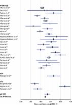
            Meta-analysis and review: effectiveness, safety, and central port design of the intraocular collamer lens

            The purpose of this review is to summarize relevant data from publications appearing in the peer-reviewed scientific literature over the past decade since US Food and Drug Administration approval of the implantable collamer lens (ICL), and, in particular, to review studies relating to sizing methodology, safety, and effectiveness, as well as more recent studies reporting clinical outcomes of the V4c Visian ICL with KS Aquaport, VICMO. A literature search was conducted using two databases, PubMed.gov and Science.gov, to identify all articles published after 2005 related to the Visian ICL (STAAR Surgical, Inc.). Articles were examined for their relevance to sizing methodology, clinical safety, and effectiveness, and the references cited in each article were also searched for additional relevant publications. The literature review revealed that all currently reported methods of determining the best-fit size of the ICL achieve similarly satisfactory results in terms of vault, the safe distance between the crystalline lens and the ICL. Specifically, meta-analysis demonstrated that sulcus-to-sulcus and white-to-white measurement-based sizing methods do not result in clinically meaningful nor statistically significant differences in vault (two-sample two-sided t-test using pooled mean and standard deviations; t (2,594)=1.33; P=0.18). The reported rates of complications related to vault are very low, except in two case series where additional risk factors such as higher levels of myopia and older age impacted the incidence of cataract. On the basis of preclinical studies and initial clinical reports, with up to 5 years of follow-up, the new VICMO central port design holds promise for further reduction of complications. Given its safety record and the significant improvement in vision and quality of life that the ICL makes possible, the benefits of ICL implantation outweigh the risks.
              Bookmark
              • Record: found
              • Abstract: found
              • Article: not found

              The development of an instrument to measure quality of vision: the Quality of Vision (QoV) questionnaire.

              To develop an instrument to measure subjective quality of vision: the Quality of Vision (QoV) questionnaire. A 30-item instrument was designed with 10 symptoms rated in each of three scales (frequency, severity, and bothersome). The QoV was completed by 900 subjects in groups of spectacle wearers, contact lens wearers, and those having had laser refractive surgery, intraocular refractive surgery, or eye disease and investigated with Rasch analysis and traditional statistics. Validity and reliability were assessed by Rasch fit statistics, principal components analysis (PCA), person separation, differential item functioning (DIF), item targeting, construct validity (correlation with visual acuity, contrast sensitivity, total root mean square [RMS] higher order aberrations [HOA]), and test-retest reliability (two-way random intraclass correlation coefficients [ICC] and 95% repeatability coefficients [R(c)]). Rasch analysis demonstrated good precision, reliability, and internal consistency for all three scales (mean square infit and outfit within 0.81-1.27; PCA >60% variance explained by the principal component; person separation 2.08, 2.10, and 2.01 respectively; and minimal DIF). Construct validity was indicated by strong correlations with visual acuity, contrast sensitivity and RMS HOA. Test-retest reliability was evidenced by a minimum ICC of 0.867 and a minimum 95% R(c) of 1.55 units. The QoV Questionnaire consists of a Rasch-tested, linear-scaled, 30-item instrument on three scales providing a QoV score in terms of symptom frequency, severity, and bothersome. It is suitable for measuring QoV in patients with all types of refractive correction, eye surgery, and eye disease that cause QoV problems.
                Bookmark

                Author and article information

                Journal
                Clin Ophthalmol
                Clin Ophthalmol
                Clinical Ophthalmology
                Clinical Ophthalmology (Auckland, N.Z.)
                Dove Medical Press
                1177-5467
                1177-5483
                2018
                27 November 2018
                : 12
                : 2427-2438
                Affiliations
                Mark Packer MD Consulting, Inc., Boulder, CO, USA, mark@ 123456markpackerconsulting.com
                Author notes
                Correspondence: Mark Packer, Mark Packer MD Consulting Inc., 1400 Bluebell Ave., Boulder, CO 80302, USA, Tel +1 541 915 0291, Fax +1 888 840 9392, Email mark@ 123456markpackerconsulting.com
                Article
                opth-12-2427
                10.2147/OPTH.S188785
                6267497
                30568421
                3a10e3d1-025a-4d80-a912-5acff8dbe9e5
                © 2018 Packer. This work is published and licensed by Dove Medical Press Limited

                The full terms of this license are available at https://www.dovepress.com/terms.php and incorporate the Creative Commons Attribution – Non Commercial (unported, v3.0) License ( http://creativecommons.org/licenses/by-nc/3.0/). By accessing the work you hereby accept the Terms. Non-commercial uses of the work are permitted without any further permission from Dove Medical Press Limited, provided the work is properly attributed.

                History
                Categories
                Review

                Ophthalmology & Optometry
                myopia,astigmatism,hyperopia,phakic refractive lens
                Ophthalmology & Optometry
                myopia, astigmatism, hyperopia, phakic refractive lens

                Comments

                Comment on this article