53
views
0
recommends
+1 Recommend
0 collections
    0
    shares
      • Record: found
      • Abstract: found
      • Article: found
      Is Open Access

      COX5B Regulates MAVS-mediated Antiviral Signaling through Interaction with ATG5 and Repressing ROS Production

      research-article

      Read this article at

      Bookmark
          There is no author summary for this article yet. Authors can add summaries to their articles on ScienceOpen to make them more accessible to a non-specialist audience.

          Abstract

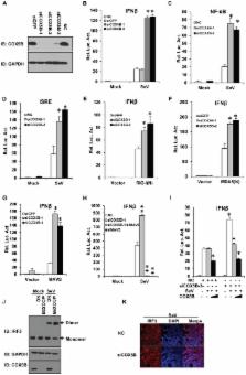
          Innate antiviral immunity is the first line of the host defense system that rapidly detects invading viruses. Mitochondria function as platforms for innate antiviral signal transduction in mammals through the adaptor protein, MAVS. Excessive activation of MAVS-mediated antiviral signaling leads to dysfunction of mitochondria and cell apoptosis that likely causes the pathogenesis of autoimmunity. However, the mechanism of how MAVS is regulated at mitochondria remains unknown. Here we show that the Cytochrome c Oxidase (CcO) complex subunit COX5B physically interacts with MAVS and negatively regulates the MAVS-mediated antiviral pathway. Mechanistically, we find that while activation of MAVS leads to increased ROS production and COX5B expression, COX5B down-regulated MAVS signaling by repressing ROS production. Importantly, our study reveals that COX5B coordinates with the autophagy pathway to control MAVS aggregation, thereby balancing the antiviral signaling activity. Thus, our study provides novel insights into the link between mitochondrial electron transport system and the autophagy pathway in regulating innate antiviral immunity.

          Author Summary

          Pattern recognition receptors are vital to innate immunity. In the antiviral innate immunity, retinoic acid-inducible gene-I (RIG-I)-like receptors (RLRs), such as RIG-I and MDA5, sense viral RNAs through their C-terminal helicase domains, then initiate the antiviral response through interaction with the essential adaptor protein MAVS, which is located in mitochondrial outer membrane. Although cumulative studies have showed that mitochondria-associated MAVS plays an important role in antiviral signaling, much remains unknown about the mechanism of MAVS activity related to mitochondrial membrane localization. In this article we demonstrate that the CcO complex subunit COX5B negatively regulates the MAVS-mediated antiviral pathway through interaction with MAVS. At the mechanistic level, we show that COX5B inhibits MAVS-mediated antiviral pathway by suppressing ROS production, and coordinating with the autophagy pathway to control MAVS aggregation. Our data support a notion that mitochondrial electron transport system coordinates with the autophagy pathway to regulate MAVS-mediated signaling for a tight control of innate antiviral immunity.

          Related collections

          Most cited references36

          • Record: found
          • Abstract: found
          • Article: not found

          Type I interferons (alpha/beta) in immunity and autoimmunity.

          The significance of type I interferons (IFN-alpha/beta) in biology and medicine renders research on their activities continuously relevant to our understanding of normal and abnormal (auto) immune responses. This relevance is bolstered by discoveries that unambiguously establish IFN-alpha/beta, among the multitude of cytokines, as dominant in defining qualitative and quantitative characteristics of innate and adaptive immune processes. Recent advances elucidating the biology of these key cytokines include better definition of their complex signaling pathways, determination of their importance in modifying the effects of other cytokines, the role of Toll-like receptors in their induction, their major cellular producers, and their broad and diverse impact on both cellular and humoral immune responses. Consequently, the role of IFN-alpha/beta in the pathogenesis of autoimmunity remains at the forefront of scientific inquiry and has begun to illuminate the mechanisms by which these molecules promote or inhibit systemic and organ-specific autoimmune diseases.
            Bookmark
            • Record: found
            • Abstract: found
            • Article: not found

            Atg9a controls dsDNA-driven dynamic translocation of STING and the innate immune response.

            Microbial nucleic acids are critical for the induction of innate immune responses, a host defense mechanism against infection by microbes. Recent studies have indicated that double-stranded DNA (dsDNA) induces potent innate immune responses via the induction of type I IFN (IFN) and IFN-inducible genes. However, the regulatory mechanisms underlying dsDNA-triggered signaling are not fully understood. Here we show that the translocation and assembly of the essential signal transducers, stimulator of IFN genes (STING) and TANK-binding kinase 1 (TBK1), are required for dsDNA-triggered innate immune responses. After sensing dsDNA, STING moves from the endoplasmic reticulum (ER) to the Golgi apparatus and finally reaches the cytoplasmic punctate structures to assemble with TBK1. The addition of an ER-retention signal to the C terminus of STING dampens its ability to induce antiviral responses. We also show that STING co-localizes with the autophagy proteins, microtubule-associated protein 1 light chain 3 (LC3) and autophagy-related gene 9a (Atg9a), after dsDNA stimulation. The loss of Atg9a, but not that of another autophagy-related gene (Atg7), greatly enhances the assembly of STING and TBK1 by dsDNA, leading to aberrant activation of the innate immune response. Hence Atg9a functions as a regulator of innate immunity following dsDNA stimulation as well as an essential autophagy protein. These results demonstrate that dynamic membrane traffic mediates the sequential translocation and assembly of STING, both of which are essential processes required for maximal activation of the innate immune response triggered by dsDNA.
              Bookmark
              • Record: found
              • Abstract: found
              • Article: not found

              Autophagy-dependent viral recognition by plasmacytoid dendritic cells.

              Plasmacytoid dendritic cells (pDCs) detect viruses in the acidified endosomes by means of Toll-like receptors (TLRs). Yet, pDC responses to certain single-stranded RNA (ssRNA) viruses occur only after live viral infection. We present evidence here that the recognition of such viruses by TLR7 requires transport of cytosolic viral replication intermediates into the lysosome by the process of autophagy. In addition, autophagy was found to be required for the production of interferon-alpha by pDCs. These results support a key role for autophagy in mediating ssRNA virus detection and interferon-alpha secretion by pDCs and suggest that cytosolic replication intermediates of viruses serve as pathogen signatures recognized by TLR7.
                Bookmark

                Author and article information

                Contributors
                Role: Editor
                Journal
                PLoS Pathog
                PLoS Pathog
                plos
                plospath
                PLoS Pathogens
                Public Library of Science (San Francisco, USA )
                1553-7366
                1553-7374
                December 2012
                December 2012
                20 December 2012
                : 8
                : 12
                : e1003086
                Affiliations
                [1 ]State Key Laboratory of Biomembrane and Membrane Biotechnology, Institute of Zoology, Chinese Academy of Sciences, Chaoyang District, Beijing, P. R. China
                [2 ]State Key Laboratory of Reproductive Biology, Institute of Zoology, Chinese Academy of Sciences, Chaoyang District, Beijing, P. R. China
                Yale University School of Medicine, United States of America
                Author notes

                The authors have declared that no competing interests exist.

                Conceived and designed the experiments: YZ DC QS. Performed the experiments: YZ XS XN LS QS. Analyzed the data: YZ DC TT QS. Wrote the paper: YZ DC QS.

                Article
                PPATHOGENS-D-12-01559
                10.1371/journal.ppat.1003086
                3534373
                23308066
                384e3f1e-5d34-42b1-b01f-6ce710bdaa1a
                Copyright @ 2012

                This is an open-access article distributed under the terms of the Creative Commons Attribution License, which permits unrestricted use, distribution, and reproduction in any medium, provided the original author and source are credited.

                History
                : 29 June 2012
                : 29 October 2012
                Page count
                Pages: 16
                Funding
                This work was supported by grants from the National Basic Research Program of China (2010CB945300), National Natural Science Foundation of China (30872349), the Ministry of Agriculture of China for Transgenic Research (2009ZX08009154-006) and the Chinese Academy of Sciences (one hundred talents program). The funders had no role in study design, data collection and analysis, decision to publish, or preparation of the manuscript.
                Categories
                Research Article
                Biology

                Infectious disease & Microbiology
                Infectious disease & Microbiology

                Comments

                Comment on this article