0
views
0
recommends
+1 Recommend
0 collections
    0
    shares
      • Record: found
      • Abstract: found
      • Article: found
      Is Open Access

      Morphology of proximal and distal human semitendinosus compartments and the effects of distal tendon harvesting for anterior cruciate ligament reconstruction

      research-article

      Read this article at

      Bookmark
          There is no author summary for this article yet. Authors can add summaries to their articles on ScienceOpen to make them more accessible to a non-specialist audience.

          Abstract

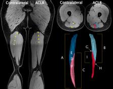
          The human semitendinosus muscle is characterized by a tendinous inscription separating proximal and distal neuromuscular compartments. As each compartment is innervated by separate nerve branches, potential exists for independent operation and control of compartments. However, the morphology and function of each compartment have not been thoroughly examined in an adult human population. Further, the distal semitendinosus tendon is typically harvested for use in anterior cruciate ligament reconstruction surgery, which induces long‐term morphological changes to the semitendinosus muscle‐tendon unit. It remains unknown if muscle morphological alterations following anterior cruciate ligament reconstruction are uniform between proximal and distal semitendinosus compartments. Here, we performed magnetic resonance imaging on 10 individuals who had undergone anterior cruciate ligament reconstruction involving an ipsilateral distal semitendinosus tendon graft 14 ± 4 months prior, extracting morphological parameters of the whole semitendinosus muscle and each individual compartment from both the (non‐injured) contralateral and surgical legs. In the contralateral leg, volume and length of the proximal compartment were smaller than the distal compartment. No between‐compartment differences in volume or length were found for anterior cruciate ligament reconstructed legs, likely due to greater shortening of the distal compared to the proximal compartment after anterior cruciate ligament reconstruction. The maximal anatomical cross‐sectional area of both compartments was substantially smaller on the anterior cruciate ligament reconstructed leg but did not differ between compartments on either leg. The absolute and relative between‐leg differences in proximal compartment morphology on the anterior cruciate ligament reconstructed leg were strongly correlated with the corresponding between‐leg differences in distal compartment morphological parameters. Specifically, greater between‐leg morphological differences in one compartment were highly correlated with large between‐leg differences in the other compartment, and vice versa for smaller differences. These relationships indicate that despite the heterogeneity in compartment length and volume, compartment atrophy is not independent or random. Further, the tendinous inscription endpoints were generally positioned at the same proximodistal level as the compartment maximal anatomical cross‐sectional areas, providing a wide area over which the tendinous inscription could mechanically interact with compartments. Overall, results suggest the two human semitendinosus compartments are not mechanically independent.

          Abstract

          Although being of anatomical intrigue for over 150 years, the human semitendinosus muscle has not been thoroughly examined in a livingadult population. Here, we used magnetic resonance imaging to describe the gross morphology of the two semitendinosus neuromuscular compartments and how each compartment may adapt to distal tendon harvesting for anterior cruciate ligament reconstruction. Despite between‐compartment differences in volume and length in non‐reconstructed legs, we found compartment morphology and adaptations are associated and not random, suggesting the two neuromuscular compartments of the human semitendinosus are not mechanically independent.

          Related collections

          Most cited references91

          • Record: found
          • Abstract: found
          • Article: not found

          Muscle volume is a major determinant of joint torque in humans.

          Muscle force (MF) is linearly related to physiological cross-sectional area (CSA), which is obtained from muscle volume (MV) divided by fibre length. Taking into account the fact that joint torque (TQ) is determined by MF multiplied by the moment arm, the maximal TQ would be a function of MV. This proposition was tested in the present study by investigating the relationship between MV and TQ for elbow flexor (EF) and extensor (EE) muscles of 26 males. The MVs of EF and EE were determined from a series of muscle CSA by magnetic resonance imaging (MRI), and pennation angle (theta) and FL by ultrasonography (US). Maximal isometric TQ was measured at right angle of elbow joint for EF and EE. There was a highly significant correlation between MV and TQ both for EF and EE (r=0.95 and 0.96 respectively) compared with that between muscle CSA and TQ, suggesting the dependence of TQ on MV. Furthermore, prediction equations for MV (MVULT) from muscle thickness (MT) measured by US was developed with reference to MVMRI by the MRI on 26 subjects, and the equations were applied to estimate MV of healthy university students (CON; 160 males) and sports athletes (ATH; 99 males). There were significant linear relationships between MVULT and TQ both for EF (r=0.783) and EE (r=0.695) for all subjects (n=259). The MVULT was significantly higher in ATH (by 32% for EF and 33% for EE, respectively) than in CON. Similarly, significantly greater TQ was observed in ATH (by 35% for EF, 37% for EE, respectively). The theta for EE showed no difference between both groups (17.8 degrees for CON and 17.5 degrees for ATH). On the other hand, the TQ to MV ratio were identical for CON and ATH. The results reveal that the muscle volume of the upper arm is a major determinant of joint torque (TQ), regardless of athletic training.
            Bookmark
            • Record: found
            • Abstract: found
            • Article: not found

            Hamstring muscles: architecture and innervation.

            Knowledge of the anatomical organization of the hamstring muscles is necessary to understand their functions, and to assist in the development of accurate clinical and biomechanical models. The hamstring muscles were examined by dissection in six embalmed human lower limbs with the purpose of clarifying their gross morphology. In addition to obtaining evidence for or against anatomical partitioning (as based on muscle architecture and pattern of innervation), data pertaining to architectural parameters such as fascicular length, volume, physiological cross-sectional area, and tendon length were collected. For each muscle, relatively consistent patterns of innervation were identified between specimens, and each was unique with respect to anatomical organization. On the basis of muscle architecture, three regions were identified within semimembranosus. However, this was not completely congruent with the pattern of innervation, as a primary nerve branch supplied only two regions, with the third region receiving a secondary branch. Semitendinosus comprised two distinct partitions arranged in series that were divided by a tendinous inscription. A singular muscle nerve or a primary nerve branch innervated each partition. In the biceps femoris long head the two regions were supplied via a primary nerve branch which divided into two primary branches or split into a series of branches. Being the only muscle to cross a single joint, biceps femoris short head consisted of two distinct regions demarcated by fiber direction, with each innervated by a separate muscle nerve. Architecturally, each muscle differed with respect to parameters such as physiological cross-sectional area, fascicular length and volume, but generally all partitions within an individual muscle were similar in fascicular length. The long proximal and distal tendons of these muscles extended into the muscle bellies thereby forming elongated musculotendinous junctions.
              Bookmark
              • Record: found
              • Abstract: found
              • Article: not found

              Muscle architecture of the human lower limb.

              The architectural features of the major knee extensors and flexors and ankle plantar flexors and dorsiflexors were determined in three human cadavers. There was marked uniformity of fiber length throughout a given muscle and a trend toward similar fiber lengths within muscles of a synergistic group. Muscle length/fiber length ratios were remarkably similar for all three limbs. Angles of fiber pinnation were relatively small (0 degree-15 degrees) and generally consistent throughout the muscle. From these architectural data, the performance of a muscle was studied with respect to its tension production and velocity of shortening potentials. The tension is a function of the number of sarcomeres in parallel, and the velocity of shortening is a function of the number of sarcomeres in series. Muscles were grouped according to whether they showed a predilection for tension or velocity of shortening.
                Bookmark

                Author and article information

                Contributors
                adam.kositsky@uef.fi
                Journal
                J Anat
                J Anat
                10.1111/(ISSN)1469-7580
                JOA
                Journal of Anatomy
                John Wiley and Sons Inc. (Hoboken )
                0021-8782
                1469-7580
                13 April 2023
                August 2023
                13 April 2023
                : 243
                : 2 , Special Section: Mini‐symposium on Vision and Visualisation ( doiID: 10.1111/joa.v243.2 )
                : 297-310
                Affiliations
                [ 1 ] Griffith Centre of Biomedical and Rehabilitation Engineering (GCORE), Menzies Health Institute Queensland Griffith University Gold Coast Queensland Australia
                [ 2 ] Department of Technical Physics University of Eastern Finland Kuopio Finland
                [ 3 ] Department of Human Movement Sciences, Faculty of Behavioural and Movement Sciences, Amsterdam Movement Sciences Vrije Universiteit Amsterdam Amsterdam The Netherlands
                [ 4 ] Mermaid Beach Radiology Gold Coast Queensland Australia
                [ 5 ] Knee Research Australia Gold Coast Queensland Australia
                Author notes
                [*] [* ] Correspondence

                Department of Technical Physics, University of Eastern Finland, Kuopio.

                Email: adam.kositsky@ 123456uef.fi

                Author information
                https://orcid.org/0000-0001-9722-6628
                https://orcid.org/0000-0002-2304-2735
                https://orcid.org/0000-0002-1784-1629
                https://orcid.org/0009-0002-1410-9843
                https://orcid.org/0000-0002-7705-9188
                https://orcid.org/0000-0002-3486-7855
                https://orcid.org/0000-0002-8447-8399
                https://orcid.org/0000-0002-2197-1856
                https://orcid.org/0000-0002-0874-7518
                Article
                JOA13869 JANAT-2022-0268.R1
                10.1111/joa.13869
                10335379
                37057314
                382fb39a-d593-4962-bdae-a0f92c24bfca
                © 2023 The Authors. Journal of Anatomy published by John Wiley & Sons Ltd on behalf of Anatomical Society.

                This is an open access article under the terms of the http://creativecommons.org/licenses/by/4.0/ License, which permits use, distribution and reproduction in any medium, provided the original work is properly cited.

                History
                : 03 March 2023
                : 24 July 2022
                : 14 March 2023
                Page count
                Figures: 6, Tables: 3, Pages: 14, Words: 10233
                Funding
                Funded by: Academy of Finland , doi 10.13039/501100002341;
                Award ID: 324529
                Award ID: 332915
                Funded by: Erasmus+ Progamme of the European Union
                Funded by: Griffith University , doi 10.13039/501100001791;
                Categories
                Original Article
                Original Articles
                Custom metadata
                2.0
                August 2023
                Converter:WILEY_ML3GV2_TO_JATSPMC version:6.3.1 mode:remove_FC converted:11.07.2023

                Anatomy & Physiology
                graft,hamstrings,magnetic resonance imaging,tendinous inscription,tenotomy
                Anatomy & Physiology
                graft, hamstrings, magnetic resonance imaging, tendinous inscription, tenotomy

                Comments

                Comment on this article