42
views
0
recommends
+1 Recommend
0 collections
    0
    shares
      • Record: found
      • Abstract: found
      • Article: found
      Is Open Access

      Prediction model for aneuploidy in early human embryo development revealed by single-cell analysis

      research-article

      Read this article at

      Bookmark
          There is no author summary for this article yet. Authors can add summaries to their articles on ScienceOpen to make them more accessible to a non-specialist audience.

          Abstract

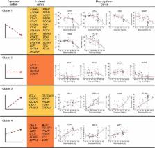
          Aneuploidies are prevalent in the human embryo and impair proper development, leading to cell cycle arrest. Recent advances in imaging and molecular and genetic analyses are postulated as promising strategies to unveil the mechanisms involved in aneuploidy generation. Here we combine time-lapse, complete chromosomal assessment and single-cell RT–qPCR to simultaneously obtain information from all cells that compose a human embryo until the approximately eight-cell stage ( n=85). Our data indicate that the chromosomal status of aneuploid embryos ( n=26), including those that are mosaic ( n=3), correlates with significant differences in the duration of the first mitotic phase when compared with euploid embryos ( n=28). Moreover, gene expression profiling suggests that a subset of genes is differentially expressed in aneuploid embryos during the first 30 h of development. Thus, we propose that the chromosomal fate of an embryo is likely determined as early as the pronuclear stage and may be predicted by a 12-gene transcriptomic signature.

          Abstract

          Aneuploidy may be fatal for the embryo, hence predicting its occurrence is important for successful in vitro fertilization. Here the authors monitor development of human preimplantation embryos in real-time and correlate the blastomere ploidy with cleavage dynamics and gene expression, identifying 12-transcript signature that determines ploidy.

          Related collections

          Most cited references57

          • Record: found
          • Abstract: found
          • Article: not found

          NIH Image to ImageJ: 25 years of image analysis.

          For the past 25 years NIH Image and ImageJ software have been pioneers as open tools for the analysis of scientific images. We discuss the origins, challenges and solutions of these two programs, and how their history can serve to advise and inform other software projects.
            Bookmark
            • Record: found
            • Abstract: found
            • Article: not found

            Genetic programs in human and mouse early embryos revealed by single-cell RNA sequencing.

            Mammalian pre-implantation development is a complex process involving dramatic changes in the transcriptional architecture. We report here a comprehensive analysis of transcriptome dynamics from oocyte to morula in both human and mouse embryos, using single-cell RNA sequencing. Based on single-nucleotide variants in human blastomere messenger RNAs and paternal-specific single-nucleotide polymorphisms, we identify novel stage-specific monoallelic expression patterns for a significant portion of polymorphic gene transcripts (25 to 53%). By weighted gene co-expression network analysis, we find that each developmental stage can be delineated concisely by a small number of functional modules of co-expressed genes. This result indicates a sequential order of transcriptional changes in pathways of cell cycle, gene regulation, translation and metabolism, acting in a step-wise fashion from cleavage to morula. Cross-species comparisons with mouse pre-implantation embryos reveal that the majority of human stage-specific modules (7 out of 9) are notably preserved, but developmental specificity and timing differ between human and mouse. Furthermore, we identify conserved key members (or hub genes) of the human and mouse networks. These genes represent novel candidates that are likely to be key in driving mammalian pre-implantation development. Together, the results provide a valuable resource to dissect gene regulatory mechanisms underlying progressive development of early mammalian embryos.
              Bookmark
              • Record: found
              • Abstract: found
              • Article: not found

              Resolution of cell fate decisions revealed by single-cell gene expression analysis from zygote to blastocyst.

              Three distinct cell types are present within the 64-cell stage mouse blastocyst. We have investigated cellular development up to this stage using single-cell expression analysis of more than 500 cells. The 48 genes analyzed were selected in part based on a whole-embryo analysis of more than 800 transcription factors. We show that in the morula, blastomeres coexpress transcription factors specific to different lineages, but by the 64-cell stage three cell types can be clearly distinguished according to their quantitative expression profiles. We identify Id2 and Sox2 as the earliest markers of outer and inner cells, respectively. This is followed by an inverse correlation in expression for the receptor-ligand pair Fgfr2/Fgf4 in the early inner cell mass. Position and signaling events appear to precede the maturation of the transcriptional program. These results illustrate the power of single-cell expression analysis to provide insight into developmental mechanisms. The technique should be widely applicable to other biological systems. Copyright 2010 Elsevier Inc. All rights reserved.
                Bookmark

                Author and article information

                Journal
                Nat Commun
                Nat Commun
                Nature Communications
                Nature Pub. Group
                2041-1723
                07 July 2015
                2015
                : 6
                : 7601
                Affiliations
                [1 ]Institute for Stem Cell Biology and Regenerative Medicine, Center for Reproductive and Stem Cell Biology , Stanford, California 94305, USA.
                [2 ]Department of Obstetrics and Gynecology, Institute for Stem Cell Biology and Regenerative Medicine, Center for Reproductive and Stem Cell Biology , Stanford, California 94305, USA.
                [3 ]IGenomix, Parc Cientific Universitat de Valencia , Catedratico Agustin Escardino 9, Paterna 46980, Valencia, Spain.
                [4 ]Department of Obstetrics and Gynecology, Fundación Instituto Valenciano de Infertilidad, University of Valencia, INCLIVA Health Research Institute , Paterna 46980, Valencia, Spain.
                Author notes
                [*]

                Present address: Division of Reproductive and Developmental Sciences, Oregon National Primate Research Center, Beaverton, Oregon 97006, USA. Department of Obstetrics and Gynecology, Oregon Health and Science University, Beaverton, Oregon 97006, USA or Department Physiology and Pharmacology, Oregon Health and Science University, Beaverton, Oregon 97006, USA

                [†]

                Present address: Department of Cell Biology and Neurosciences, Montana State University, Bozeman, Montana 59717, USA or Department of Chemistry and Biochemistry, Montana State University, Bozeman, Montana 59717, USA

                Article
                ncomms8601
                10.1038/ncomms8601
                4506544
                26151134
                3754c5dc-3b89-4062-a954-79e4438376d2
                Copyright © 2015, Nature Publishing Group, a division of Macmillan Publishers Limited. All Rights Reserved.

                This work is licensed under a Creative Commons Attribution 4.0 International License. The images or other third party material in this article are included in the article's Creative Commons license, unless indicated otherwise in the credit line; if the material is not included under the Creative Commons license, users will need to obtain permission from the license holder to reproduce the material. To view a copy of this license, visit http://creativecommons.org/licenses/by/4.0/

                History
                : 26 September 2014
                : 22 May 2015
                Categories
                Article

                Uncategorized
                Uncategorized

                Comments

                Comment on this article