51
views
0
recommends
+1 Recommend
0 collections
    0
    shares
      • Record: found
      • Abstract: found
      • Article: found
      Is Open Access

      Establishment of a Human Blood-Brain Barrier Co-culture Model Mimicking the Neurovascular Unit Using Induced Pluri- and Multipotent Stem Cells

      research-article

      Read this article at

      Bookmark
          There is no author summary for this article yet. Authors can add summaries to their articles on ScienceOpen to make them more accessible to a non-specialist audience.

          Summary

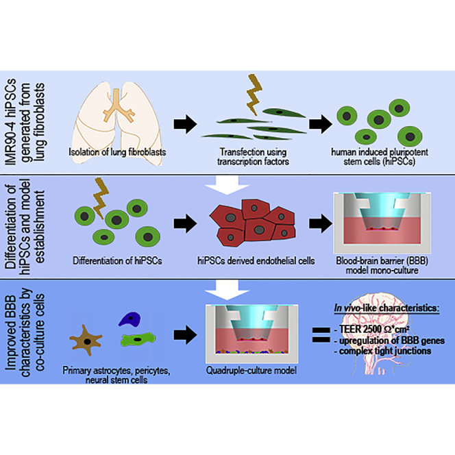
          In vitro models of the human blood-brain barrier (BBB) are highly desirable for drug development. This study aims to analyze a set of ten different BBB culture models based on primary cells, human induced pluripotent stem cells (hiPSCs), and multipotent fetal neural stem cells (fNSCs). We systematically investigated the impact of astrocytes, pericytes, and NSCs on hiPSC-derived BBB endothelial cell function and gene expression. The quadruple culture models, based on these four cell types, achieved BBB characteristics including transendothelial electrical resistance (TEER) up to 2,500 Ω cm 2 and distinct upregulation of typical BBB genes. A complex in vivo-like tight junction (TJ) network was detected by freeze-fracture and transmission electron microscopy. Treatment with claudin-specific TJ modulators caused TEER decrease, confirming the relevant role of claudin subtypes for paracellular tightness. Drug permeability tests with reference substances were performed and confirmed the suitability of the models for drug transport studies.

          Graphical Abstract

          Highlights

          • Establishment of a standardized human BBB co-culture model based on hiPSCs and fNSCs

          • Reflection of physiological BBB integrity and expression of relevant transporters/TJs

          • Confirmation of TJ network functionality by claudin-specific TJ modulators

          • Validation of physiological transcellular model tightness by permeability studies

          Abstract

          In this article, Metzger and colleagues present the establishment of physiologically relevant human blood-brain barrier quadruple culture models based on induced pluripotent and multipotent stem cells. The novel model can be used as a powerful tool in pharmaceutical drug research and ADMET studies, which may allow a more reliable translation of promising drug candidates into clinical application.

          Related collections

          Most cited references37

          • Record: found
          • Abstract: found
          • Article: not found

          Electrical resistance across the blood-brain barrier in anaesthetized rats: a developmental study.

          1. Ion permeability of the blood-brain barrier was studied by in situ measurement of transendothelial electrical resistance in anaesthetized rats aged between 17 days gestation and 33 days after birth, and by electron microscopic examination of lanthanum permeability in fetal and neonatal rats aged up to 10 days old. 2. The blood-brain barrier in 17- to 20-day fetuses had a resistance of 310 omega cm2 but was impermeable to lanthanum, and therefore had properties intermediate between leaky and tight epithelia. 3. From 21 days gestation, the resistance was 1128 omega cm2, indicating a tight blood-brain barrier and low ion permeability. There was little further change in barrier resistance after birth, and in 28- to 33-day rats, when the brain barrier systems are mature in other ways, vessels had a mean resistance of 1462 omega cm2. 4. In the tight blood-brain barrier, arterial vessels had a significantly higher resistance than venous vessels, 1490 and 918 omega cm2 respectively. In vessels less than 50 microns diameter and within the normal 60 min experimental period, there was no significant variation in vessel resistance. 5. Hyperosmotic shock caused a rapid decay in resistance (maximal within 5 min), and after disruption of the blood-brain barrier, vessel resistance was 100-300 omega cm2 in both arterial and venous vessels, and the effect was reversible. After the application of metabolic poisons (NaCN plus iodoacetate) and low temperature there was a similarly low electrical resistance. 6. It is concluded that the increase in electrical resistance at birth indicates a decrease in paracellular ion permeability at the blood-brain barrier and is required for effective brain interstitial fluid ion regulation.
            Bookmark
            • Record: found
            • Abstract: found
            • Article: not found

            Permeability studies on in vitro blood-brain barrier models: physiology, pathology, and pharmacology.

            (1) The specifically regulated restrictive permeability barrier to cells and molecules is the most important feature of the blood-brain barrier (BBB). The aim of this review was to summarize permeability data obtained on in vitro BBB models by measurement of transendothelial electrical resistance and by calculation of permeability coefficients for paracellular or transendothelial tracers. (2) Results from primary cultures of cerebral microvascular endothelial cells or immortalized cell lines from bovine, human, porcine, and rodent origin are presented. Effects of coculture with astroglia, neurons, mesenchymal cells, blood cells, and conditioned media, as well as physiological influence of serum components, hormones, growth factors, lipids, and lipoproteins on the barrier function are discussed. (3) BBB permeability results gained on in vitro models of pathological conditions including hypoxia and reoxygenation, neurodegenerative diseases, or bacterial and viral infections have been reviewed. Effects of cytokines, vasoactive mediators, and other pathogenic factors on barrier integrity are also detailed. (4) Pharmacological treatments modulating intracellular cyclic nucleotide or calcium levels, and activity of protein kinases, protein tyrosine phosphatases, phospholipases, cyclooxygenases, or lipoxygenases able to change BBB integrity are outlined. Barrier regulation by drugs involved in the metabolism of nitric oxide and reactive oxygen species, as well as influence of miscellaneous treatments are also listed and evaluated. (5) Though recent advances resulted in development of improved in vitro BBB model systems to investigate disease modeling, drug screening, and testing vectors targeting the brain, there is a need for checking validity of permeability models and cautious interpretation of data.
              Bookmark
              • Record: found
              • Abstract: found
              • Article: found
              Is Open Access

              Comparative study of four immortalized human brain capillary endothelial cell lines, hCMEC/D3, hBMEC, TY10, and BB19, and optimization of culture conditions, for an in vitro blood–brain barrier model for drug permeability studies

              Background Reliable human in vitro blood–brain barrier (BBB) models suitable for high-throughput screening are urgently needed in early drug discovery and development for assessing the ability of promising bioactive compounds to overcome the BBB. To establish an improved human in vitro BBB model, we compared four currently available and well characterized immortalized human brain capillary endothelial cell lines, hCMEC/D3, hBMEC, TY10, and BB19, with respect to barrier tightness and paracellular permeability. Co-culture systems using immortalized human astrocytes (SVG-A cell line) and immortalized human pericytes (HBPCT cell line) were designed with the aim of positively influencing barrier tightness. Methods Tight junction (TJ) formation was assessed by transendothelial electrical resistance (TEER) measurements using a conventional epithelial voltohmmeter (EVOM) and an automated CellZscope system which records TEER and cell layer capacitance (CCL) in real-time. Paracellular permeability was assessed using two fluorescent marker compounds with low BBB penetration (sodium fluorescein (Na-F) and lucifer yellow (LY)). Conditions were optimized for each endothelial cell line by screening a series of 24-well tissue culture inserts from different providers. For hBMEC cells, further optimization was carried out by varying coating material, coating procedure, cell seeding density, and growth media composition. Biochemical characterization of cell type-specific transmembrane adherens junction protein VE-cadherin and of TJ proteins ZO-1 and claudin-5 were carried out for each endothelial cell line. In addition, immunostaining for ZO-1 in hBMEC cell line was performed. Results The four cell lines all expressed the endothelial cell type-specific adherens junction protein VE-cadherin. The TJ protein ZO-1 was expressed in hCMEC/D3 and in hBMEC cells. ZO-1 expression could be confirmed in hBMEC cells by immunocytochemical staining. Claudin-5 expression was detected in hCMEC/D3, TY10, and at a very low level in hBMEC cells. Highest TEER values and lowest paracellular permeability for Na-F and LY were obtained with mono-cultures of hBMEC cell line when cultivated on 24-well tissue culture inserts from Greiner Bio-one® (transparent PET membrane, 3.0 μm pore size). In co-culture models with SVG-A and HBPCT cells, no increase of TEER could be observed, suggesting that none of the investigated endothelial cell lines responded positively to stimuli from immortalized astrocytic or pericytic cells. Conclusions Under the conditions examined in our experiments, hBMEC proved to be the most suitable human cell line for an in vitro BBB model concerning barrier tightness in a 24-well mono-culture system intended for higher throughput. This BBB model is being validated with several compounds (known to cross or not to cross the BBB), and will potentially be selected for the assessment of BBB permeation of bioactive natural products.
                Bookmark

                Author and article information

                Contributors
                Journal
                Stem Cell Reports
                Stem Cell Reports
                Stem Cell Reports
                Elsevier
                2213-6711
                23 March 2017
                11 April 2017
                23 March 2017
                : 8
                : 4
                : 894-906
                Affiliations
                [1 ]University Hospital Würzburg, Chair Tissue Engineering and Regenerative Medicine, 97070 Würzburg, Germany
                [2 ]Translational Center Würzburg “Regenerative Therapies for Oncology and Musculoskeletal Diseases”, Branch of Fraunhofer Institute for Interfacial Engineering and Biotechnology IGB, 97070 Würzburg, Germany
                [3 ]Julius-Maximilians-University Würzburg, Institute of Anatomy and Cell Biology, Stem Cell and Regenerative Medicine Group, 97070 Würzburg, Germany
                [4 ]Leopold-Franzens-University Innsbruck, Institute of Molecular Biology & CMBI, Department Genomics, Stem Cell Biology & Regenerative Medicine, 6020 Innsbruck, Austria
                [5 ]Charité Universitätsmedizin Berlin, Clinical Physiology & Nutritional Medicine, Department of Gastroenterology, Rheumatology & Infectious Diseases, 12203 Berlin, Germany
                [6 ]Leibniz Institut für Molekulare Pharmakologie, 13125 Berlin, Germany
                [7 ]University Hospital Würzburg, Women’s Hospital and Polyclinic, 97080 Würzburg, Germany
                [8 ]AIT Austrian Institute of Technology GmbH, Competence Center Health and Bioresources, Competence Unit Molecular Diagnostics, 1190 Vienna, Austria
                Author notes
                []Corresponding author marco.metzger@ 123456igb.fraunhofer.de
                [9]

                Co-senior author

                Article
                S2213-6711(17)30088-7
                10.1016/j.stemcr.2017.02.021
                5390136
                28344002
                3732c8b7-4f95-4f1f-940d-71afe32e44d3
                © 2017 The Author(s)

                This is an open access article under the CC BY-NC-ND license (http://creativecommons.org/licenses/by-nc-nd/4.0/).

                History
                : 8 April 2016
                : 22 February 2017
                : 23 February 2017
                Categories
                Article

                blood-brain barrier (bbb) model,human induced pluripotent stem cells (hipscs),multipotent fetal neural stem cells (fnscs),neurovascular unit in vitro

                Comments

                Comment on this article