10
views
0
recommends
+1 Recommend
0 collections
    0
    shares
      • Record: found
      • Abstract: found
      • Article: found
      Is Open Access

      Papillary lesions of the breast

      review-article

      Read this article at

      Bookmark
          There is no author summary for this article yet. Authors can add summaries to their articles on ScienceOpen to make them more accessible to a non-specialist audience.

          Abstract

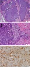
          Papillary lesions of the breast represent a heterogeneous group of lesions including benign papillomas, papillomas with focal epithelial atypia, fully fledged ductal carcinoma in situ (DCIS) or lobular neoplasia, papillary DCIS, encapsulated papillary carcinomas without or with invasion, solid papillary carcinomas, and invasive papillary carcinomas. A micropapillary pattern characterized by lack of fibrous stalks within the papillae is observed in micropapillary DCIS and invasive micropapillary carcinoma. In addition, a variety of other rare breast lesions reveals a papillary architecture such as tall cell carcinoma with reversed polarity (TCCRP) and mucinous cystadenocarcinoma, adenomyoepithelioma, and secretory carcinoma. In addition, benign lesions such as usual ductal hyperplasia, apocrine metaplasia, gynecomastia, and juvenile papillomatosis may show a papillary or micropapillary architecture. Fragments of a benign papilloma in a breast biopsy are considered a lesion of uncertain malignant potential (B3 in the European classification) and excision is mostly recommended. Although the knowledge about molecular pathology of papillary breast lesions has increased, there is not sufficient evidence for diagnostically useful molecular features, yet. The aim of this review is to provide an update on papillary and micropapillary lesions with emphasis on problematic areas for daily diagnostic work including biopsies.

          Related collections

          Most cited references80

          • Record: found
          • Abstract: found
          • Article: not found

          Expression of the ETV6-NTRK3 gene fusion as a primary event in human secretory breast carcinoma.

          We report that human secretory breast carcinoma (SBC), a rare subtype of infiltrating ductal carcinoma, expresses the ETV6-NTRK3 gene fusion previously cloned in pediatric mesenchymal cancers. This gene fusion encodes a chimeric tyrosine kinase with potent transforming activity in fibroblasts. ETV6-NTRK3 expression was confirmed in 12 (92%) of 13 SBC cases, but not in other ductal carcinomas. Retroviral transfer of ETV6-NTRK3 (EN) into murine mammary epithelial cells resulted in transformed cells that readily formed tumors in nude mice. Phenotypically, tumors produced glands and expressed epithelial antigens, confirming that EN transformation is compatible with epithelial differentiation. This represents a recurrent chromosomal rearrangement and expression of a dominantly acting oncogene as a primary event in human breast carcinoma.
            Bookmark
            • Record: found
            • Abstract: found
            • Article: found
            Is Open Access

            Second International Consensus Conference on lesions of uncertain malignant potential in the breast (B3 lesions)

            Purpose The second International Consensus Conference on B3 lesions was held in Zurich, Switzerland, in March 2018, organized by the International Breast Ultrasound School to re-evaluate the consensus recommendations. Methods This study (1) evaluated how management recommendations of the first Zurich Consensus Conference of 2016 on B3 lesions had influenced daily practice and (2) reviewed current literature towards recommendations to biopsy. Results In 2018, the consensus recommendations for management of B3 lesions remained almost unchanged: For flat epithelial atypia (FEA), classical lobular neoplasia (LN), papillary lesions (PL) and radial scars (RS) diagnosed on core-needle biopsy (CNB) or vacuum-assisted biopsy (VAB), excision by VAB in preference to open surgery, and for atypical ductal hyperplasia (ADH) and phyllodes tumors (PT) diagnosed at VAB or CNB, first-line open surgical excision (OE) with follow-up surveillance imaging for 5 years. Analyzing the Database of the Swiss Minimally Invasive Breast Biopsies (MIBB) with more than 30,000 procedures recorded, there was a significant increase in recommending more frequent surveillance of LN [65% in 2018 vs. 51% in 2016 (p = 0.004)], FEA (72% in 2018 vs. 62% in 2016 (p = 0.005)), and PL [(76% in 2018 vs. 70% in 2016 (p = 0.04)] diagnosed on VAB. A trend to more frequent surveillance was also noted also for RS [77% in 2018 vs. 67% in 2016 (p = 0.07)]. Conclusions Minimally invasive management of B3 lesions (except ADH and PT) with VAB continues to be appropriate as an alternative to first-line OE in most cases, but with more frequent surveillance, especially for LN.
              Bookmark
              • Record: found
              • Abstract: found
              • Article: not found

              Secretory carcinoma of the breast: results from the survival, epidemiology and end results database.

              Secretory carcinoma of the breast is a rare breast cancer that is associated with incidence at a young age and an indolent course. The role for breast conservation and adjuvant radiation is unknown. The SEER database was reviewed and 83 patients were identified with secretory carcinoma of the breast between the years 1983 and 2007. Baseline characteristics were compared with χ(2) or Fisher's exact test. Overall survival (OS) and cause-specific survival (CSS) were estimated using the Kaplan-Meier method. Analyses were performed using PASW Statistics, version 18. Median follow-up was 70 months. Median patient age was 53 years (range 11-86 years). 29 patients (34.9%) had involved regional lymph nodes. 39 patients (47.0%) underwent lumpectomy and 44 patients (53.0%) underwent mastectomy. 35 patients (42.2%) received radiation. Patients receiving radiation were more likely to have undergone lumpectomy, and the use of radiation increased over time. 5-year OS was 87.2%; 10-year OS was 76.5%. 5-year CSS was 94.4%; 10-year CSS was 91.4%. Among the lumpectomy patients, 25 patients (64.1%) received radiation. For lumpectomy patients, those who did not receive radiation had a 5-year OS of 92.9% and 10-year OS of 72.2% while patients who did receive radiation had a 5-year OS of 95.5% and 10-year OS of 85.9%. Only 1 patient treated with lumpectomy only died of cancer (92.9% CSS) and no patients treated with lumpectomy and radiation died of cancer (100% CSS). Secretory carcinoma of the breast commonly occurs at a later age than previously recognized, and is associated with good long-term survival. Copyright © 2012 Elsevier Ltd. All rights reserved.
                Bookmark

                Author and article information

                Contributors
                kulka.janina@med.semmelweis-univ.hu
                Journal
                Virchows Arch
                Virchows Arch
                Virchows Archiv
                Springer Berlin Heidelberg (Berlin/Heidelberg )
                0945-6317
                1432-2307
                3 November 2021
                3 November 2021
                2022
                : 480
                : 1
                : 65-84
                Affiliations
                [1 ]GRID grid.11804.3c, ISNI 0000 0001 0942 9821, 2nd Department of Pathology, , Semmelweis University, ; Üllői út 93, 1091 Budapest, Hungary E.U.
                [2 ]GRID grid.417105.6, ISNI 0000 0004 0621 6048, Department of Pathology, , Uzsoki Hospital, ; Budapest, Hungary
                [3 ]GRID grid.410569.f, ISNI 0000 0004 0626 3338, Department of Imaging and Pathology, Laboratory of Translational Cell & Tissue Research, KU Leuven, , University of Leuven, University Hospitals Leuven, ; Leuven, Belgium
                [4 ]Department of Pathology, Hospital Graz II, Graz, Austria
                [5 ]GRID grid.9970.7, ISNI 0000 0001 1941 5140, School of Medicine, , Johannes Kepler University, ; Linz, Austria
                Author information
                http://orcid.org/0000-0001-6498-5943
                Article
                3182
                10.1007/s00428-021-03182-7
                8983543
                34734332
                36352c02-6d1b-4324-baf3-a964336e4e91
                © The Author(s) 2021

                Open AccessThis article is licensed under a Creative Commons Attribution 4.0 International License, which permits use, sharing, adaptation, distribution and reproduction in any medium or format, as long as you give appropriate credit to the original author(s) and the source, provide a link to the Creative Commons licence, and indicate if changes were made. The images or other third party material in this article are included in the article's Creative Commons licence, unless indicated otherwise in a credit line to the material. If material is not included in the article's Creative Commons licence and your intended use is not permitted by statutory regulation or exceeds the permitted use, you will need to obtain permission directly from the copyright holder. To view a copy of this licence, visit http://creativecommons.org/licenses/by/4.0/.

                History
                : 21 May 2021
                : 1 August 2021
                : 6 August 2021
                Funding
                Funded by: Semmelweis University
                Categories
                Review
                Custom metadata
                © Springer-Verlag GmbH Germany, part of Springer Nature 2022

                Pathology
                breast,papillary lesions,ductal carcinoma in situ,dcis,micropapillary,biopsy
                Pathology
                breast, papillary lesions, ductal carcinoma in situ, dcis, micropapillary, biopsy

                Comments

                Comment on this article