0
views
0
recommends
+1 Recommend
0 collections
    0
    shares
      • Record: found
      • Abstract: found
      • Article: found
      Is Open Access

      Clinical study on the role of LncRNA STX17‐AS1 in wound healing and hypertrophic scar formation

      research-article

      Read this article at
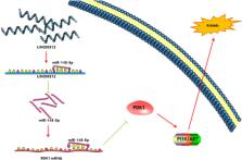
      Bookmark
          There is no author summary for this article yet. Authors can add summaries to their articles on ScienceOpen to make them more accessible to a non-specialist audience.

          Abstract

          Wound healing is a complex process that can lead to hypertrophic scarring (HS) when dysregulated. The role of lncRNAs in this process is increasingly recognized, yet the specific contributions of lncRNA STX17‐AS1 require elucidation. This study investigated the expression of STX17‐AS1, its regulatory effects on miR‐145‐5p, and downstream targets, highlighting its impact on wound repair and HS development. In a cohort of 20 HS patients and 20 matched controls, we assessed the expression of STX17‐AS1, miR‐145‐5p and PDK1 via real‐time PCR and immunohistochemistry. We correlated these expressions with wound characteristics and analysed their regulatory impact on the PI3K/AKT pathway, crucial for cellular proliferation and migration in wound healing. Elevated levels of STX17‐AS1 and miR‐145‐5p in patient samples were correlated with larger wound areas and slower healing rates, suggesting the regulatory imbalance in scar formation. The negative correlation of PDK1 expression with age and its positive association with wound size underscored its relevance in wound repair mechanisms. Functional analysis confirmed the interaction between STX17‐AS1 and miR‐145‐5p and modulation of PDK1, indicating the potential disruption of the PI3K/AKT pathway in the wound healing process. The study identified lncRNA STX17‐AS1 as the significant mediator in wound healing, with aberrations in its pathway correlating with impaired healing and HS. The findings proposed lncRNA STX17‐AS1 and miR‐145‐5p as molecular targets to enhance wound healing and prevent pathological scarring, offering a new avenue for therapeutic advances in wound management and regenerative medicine.

          Related collections

          Most cited references25

          • Record: found
          • Abstract: found
          • Article: not found

          Factors affecting wound healing.

          Wound healing, as a normal biological process in the human body, is achieved through four precisely and highly programmed phases: hemostasis, inflammation, proliferation, and remodeling. For a wound to heal successfully, all four phases must occur in the proper sequence and time frame. Many factors can interfere with one or more phases of this process, thus causing improper or impaired wound healing. This article reviews the recent literature on the most significant factors that affect cutaneous wound healing and the potential cellular and/or molecular mechanisms involved. The factors discussed include oxygenation, infection, age and sex hormones, stress, diabetes, obesity, medications, alcoholism, smoking, and nutrition. A better understanding of the influence of these factors on repair may lead to therapeutics that improve wound healing and resolve impaired wounds.
            Bookmark
            • Record: found
            • Abstract: found
            • Article: not found

            Neuronal Cell Death.

            Neuronal cell death occurs extensively during development and pathology, where it is especially important because of the limited capacity of adult neurons to proliferate or be replaced. The concept of cell death used to be simple as there were just two or three types, so we just had to work out which type was involved in our particular pathology and then block it. However, we now know that there are at least a dozen ways for neurons to die, that blocking a particular mechanism of cell death may not prevent the cell from dying, and that non-neuronal cells also contribute to neuronal death. We review here the mechanisms of neuronal death by intrinsic and extrinsic apoptosis, oncosis, necroptosis, parthanatos, ferroptosis, sarmoptosis, autophagic cell death, autosis, autolysis, paraptosis, pyroptosis, phagoptosis, and mitochondrial permeability transition. We next explore the mechanisms of neuronal death during development, and those induced by axotomy, aberrant cell-cycle reentry, glutamate (excitoxicity and oxytosis), loss of connected neurons, aggregated proteins and the unfolded protein response, oxidants, inflammation, and microglia. We then reassess which forms of cell death occur in stroke and Alzheimer's disease, two of the most important pathologies involving neuronal cell death. We also discuss why it has been so difficult to pinpoint the type of neuronal death involved, if and why the mechanism of neuronal death matters, the molecular overlap and interplay between death subroutines, and the therapeutic implications of these multiple overlapping forms of neuronal death.
              Bookmark
              • Record: found
              • Abstract: found
              • Article: found
              Is Open Access

              Fibroblasts and myofibroblasts in wound healing

              (Myo)fibroblasts are key players for maintaining skin homeostasis and for orchestrating physiological tissue repair. (Myo)fibroblasts are embedded in a sophisticated extracellular matrix (ECM) that they secrete, and a complex and interactive dialogue exists between (myo)fibroblasts and their microenvironment. In addition to the secretion of the ECM, (myo)fibroblasts, by secreting matrix metalloproteinases and tissue inhibitors of metalloproteinases, are able to remodel this ECM. (Myo)fibroblasts and their microenvironment form an evolving network during tissue repair, with reciprocal actions leading to cell differentiation, proliferation, quiescence, or apoptosis, and actions on growth factor bioavailability by binding, sequestration, and activation. In addition, the (myo)fibroblast phenotype is regulated by mechanical stresses to which they are subjected and thus by mechanical signaling. In pathological situations (excessive scarring or fibrosis), or during aging, this dialogue between the (myo)fibroblasts and their microenvironment may be altered or disrupted, leading to repair defects or to injuries with damaged and/or cosmetic skin alterations such as wrinkle development. The intimate dialogue between the (myo)fibroblasts and their microenvironment therefore represents a fascinating domain that must be better understood in order not only to characterize new therapeutic targets and drugs able to prevent or treat pathological developments but also to interfere with skin alterations observed during normal aging or premature aging induced by a deleterious environment.
                Bookmark

                Author and article information

                Contributors
                chejiangwang_tfch@163.com
                Journal
                Int Wound J
                Int Wound J
                10.1111/(ISSN)1742-481X
                IWJ
                International Wound Journal
                Blackwell Publishing Ltd (Oxford, UK )
                1742-4801
                1742-481X
                31 January 2024
                February 2024
                : 21
                : 2 ( doiID: 10.1111/iwj.v21.2 )
                : e14658
                Affiliations
                [ 1 ] Department of Orthopedic and Burn Surgery Tianjin First Central Hospital Tianjin China
                Author notes
                [*] [* ] Correspondence

                Chejiang Wang, Department of Orthopedic and Burn Surgery, Tianjin First Central Hospital, Tianjin 300190, China.

                Email: chejiangwang_tfch@ 123456163.com

                Author information
                https://orcid.org/0009-0002-9681-0616
                Article
                IWJ14658
                10.1111/iwj.14658
                10830909
                3300d247-6bc5-46ac-9f82-22c89d1b4f75
                © 2024 The Authors. International Wound Journal published by Medicalhelplines.com Inc and John Wiley & Sons Ltd.

                This is an open access article under the terms of the http://creativecommons.org/licenses/by-nc-nd/4.0/ License, which permits use and distribution in any medium, provided the original work is properly cited, the use is non‐commercial and no modifications or adaptations are made.

                History
                : 23 December 2023
                : 13 December 2023
                : 24 December 2023
                Page count
                Figures: 3, Tables: 6, Pages: 8, Words: 4036
                Funding
                Funded by: General scientific and technological projects of Tianjin Health and Wellness Committee
                Award ID: TJWJ2022MS016
                Categories
                Original Article
                Original Articles
                Custom metadata
                2.0
                February 2024
                Converter:WILEY_ML3GV2_TO_JATSPMC version:6.3.6 mode:remove_FC converted:31.01.2024

                Emergency medicine & Trauma
                hypertrophic scarring,lncrna stx17‐as1,mir‐145‐5p,pi3k/akt pathway,wound healing

                Comments

                Comment on this article