20
views
0
recommends
+1 Recommend
0 collections
    0
    shares
      • Record: found
      • Abstract: found
      • Article: found
      Is Open Access

      The High Costs of Low-Grade Inflammation: Persistent Fatigue as a Consequence of Reduced Cellular-Energy Availability and Non-adaptive Energy Expenditure

      review-article

      Read this article at

      Bookmark
          There is no author summary for this article yet. Authors can add summaries to their articles on ScienceOpen to make them more accessible to a non-specialist audience.

          Abstract

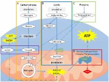
          Chronic or persistent fatigue is a common, debilitating symptom of several diseases. Persistent fatigue has been associated with low-grade inflammation in several models of fatigue, including cancer-related fatigue and chronic fatigue syndrome. However, it is unclear how low-grade inflammation leads to the experience of fatigue. We here propose a model of an imbalance in energy availability and energy expenditure as a consequence of low-grade inflammation. In this narrative review, we discuss how chronic low-grade inflammation can lead to reduced cellular-energy availability. Low-grade inflammation induces a metabolic switch from energy-efficient oxidative phosphorylation to fast-acting, but less efficient, aerobic glycolytic energy production; increases reactive oxygen species; and reduces insulin sensitivity. These effects result in reduced glucose availability and, thereby, reduced cellular energy. In addition, emerging evidence suggests that chronic low-grade inflammation is associated with increased willingness to exert effort under specific circumstances. Circadian-rhythm changes and sleep disturbances might mediate the effects of inflammation on cellular-energy availability and non-adaptive energy expenditure. In the second part of the review, we present evidence for these metabolic pathways in models of persistent fatigue, focusing on chronic fatigue syndrome and cancer-related fatigue. Most evidence for reduced cellular-energy availability in relation to fatigue comes from studies on chronic fatigue syndrome. While the mechanistic evidence from the cancer-related fatigue literature is still limited, the sparse results point to reduced cellular-energy availability as well. There is also mounting evidence that behavioral-energy expenditure exceeds the reduced cellular-energy availability in patients with persistent fatigue. This suggests that an inability to adjust energy expenditure to available resources might be one mechanism underlying persistent fatigue.

          Related collections

          Most cited references117

          • Record: found
          • Abstract: found
          • Article: not found

          Mitochondrial defects and oxidative stress in Alzheimer disease and Parkinson disease.

          Alzheimer disease (AD) and Parkinson disease (PD) are the two most common age-related neurodegenerative diseases characterized by prominent neurodegeneration in selective neural systems. Although a small fraction of AD and PD cases exhibit evidence of heritability, among which many genes have been identified, the majority are sporadic without known causes. Molecular mechanisms underlying neurodegeneration and pathogenesis of these diseases remain elusive. Convincing evidence demonstrates oxidative stress as a prominent feature in AD and PD and links oxidative stress to the development of neuronal death and neural dysfunction, which suggests a key pathogenic role for oxidative stress in both AD and PD. Notably, mitochondrial dysfunction is also a prominent feature in these diseases, which is likely to be of critical importance in the genesis and amplification of reactive oxygen species and the pathophysiology of these diseases. In this review, we focus on changes in mitochondrial DNA and mitochondrial dynamics, two aspects critical to the maintenance of mitochondrial homeostasis and function, in relationship with oxidative stress in the pathogenesis of AD and PD. Copyright © 2012 Elsevier Inc. All rights reserved.
            Bookmark
            • Record: found
            • Abstract: found
            • Article: found
            Is Open Access

            Circadian Misalignment Augments Markers of Insulin Resistance and Inflammation, Independently of Sleep Loss

            Shift workers, who are exposed to irregular sleep schedules resulting in sleep deprivation and misalignment of circadian rhythms, have an increased risk of diabetes relative to day workers. In healthy adults, sleep restriction without circadian misalignment promotes insulin resistance. To determine whether the misalignment of circadian rhythms that typically occurs in shift work involves intrinsic adverse metabolic effects independently of sleep loss, a parallel group design was used to study 26 healthy adults. Both interventions involved 3 inpatient days with 10-h bedtimes, followed by 8 inpatient days of sleep restriction to 5 h with fixed nocturnal bedtimes (circadian alignment) or with bedtimes delayed by 8.5 h on 4 of the 8 days (circadian misalignment). Daily total sleep time (SD) during the intervention was nearly identical in the aligned and misaligned conditions (4 h 48 min [5 min] vs. 4 h 45 min [6 min]). In both groups, insulin sensitivity (SI) significantly decreased after sleep restriction, without a compensatory increase in insulin secretion, and inflammation increased. In male participants exposed to circadian misalignment, the reduction in SI and the increase in inflammation both doubled compared with those who maintained regular nocturnal bedtimes. Circadian misalignment that occurs in shift work may increase diabetes risk and inflammation, independently of sleep loss.
              Bookmark
              • Record: found
              • Abstract: found
              • Article: not found

              Elevated Mitochondrial Bioenergetics and Axonal Arborization Size Are Key Contributors to the Vulnerability of Dopamine Neurons.

              Although the mechanisms underlying the loss of neurons in Parkinson's disease are not well understood, impaired mitochondrial function and pathological protein aggregation are suspected as playing a major role. Why DA (dopamine) neurons and a select small subset of brain nuclei are particularly vulnerable to such ubiquitous cellular dysfunctions is presently one of the key unanswered questions in Parkinson's disease research. One intriguing hypothesis is that their heightened vulnerability is a consequence of their elevated bioenergetic requirements. Here, we show for the first time that vulnerable nigral DA neurons differ from less vulnerable DA neurons such as those of the VTA (ventral tegmental area) by having a higher basal rate of mitochondrial OXPHOS (oxidative phosphorylation), a smaller reserve capacity, a higher density of axonal mitochondria, an elevated level of basal oxidative stress, and a considerably more complex axonal arborization. Furthermore, we demonstrate that reducing axonal arborization by acting on axon guidance pathways with Semaphorin 7A reduces in parallel the basal rate of mitochondrial OXPHOS and the vulnerability of nigral DA neurons to the neurotoxic agents MPP(+) (1-methyl-4-phenylpyridinium) and rotenone. Blocking L-type calcium channels with isradipine was protective against MPP(+) but not rotenone. Our data provide the most direct demonstration to date in favor of the hypothesis that the heightened vulnerability of nigral DA neurons in Parkinson's disease is directly due to their particular bioenergetic and morphological characteristics.
                Bookmark

                Author and article information

                Contributors
                Journal
                Front Behav Neurosci
                Front Behav Neurosci
                Front. Behav. Neurosci.
                Frontiers in Behavioral Neuroscience
                Frontiers Media S.A.
                1662-5153
                26 April 2018
                2018
                : 12
                : 78
                Affiliations
                Neuroimmunology Laboratory, Symptom Research Department, The University of Texas MD Anderson Cancer Institute , Houston, TX, United States
                Author notes

                Edited by: Martin Hadamitzky, Universitätsklinikum Essen, Germany

                Reviewed by: Andrew Chan, Ruhr University Bochum, Germany; Alessandro Laviano, Sapienza Università di Roma, Italy; Lisa Mullen, Brighton and Sussex Medical School, University of Sussex, United Kingdom

                *Correspondence: Tamara E. Lacourt tlacourt@ 123456mdanderson.org
                Article
                10.3389/fnbeh.2018.00078
                5932180
                29755330
                326ff369-f722-4c5a-8d0a-0a1c589c0142
                Copyright © 2018 Lacourt, Vichaya, Chiu, Dantzer and Heijnen.

                This is an open-access article distributed under the terms of the Creative Commons Attribution License (CC BY). The use, distribution or reproduction in other forums is permitted, provided the original author(s) and the copyright owner are credited and that the original publication in this journal is cited, in accordance with accepted academic practice. No use, distribution or reproduction is permitted which does not comply with these terms.

                History
                : 20 December 2017
                : 09 April 2018
                Page count
                Figures: 3, Tables: 3, Equations: 0, References: 143, Pages: 20, Words: 15509
                Funding
                Funded by: National Institutes of Health 10.13039/100000002
                Award ID: RO1ns073939
                Award ID: RO1CA193522
                Award ID: RO1CA208371
                Categories
                Neuroscience
                Review

                Neurosciences
                cytokines,metabolism,effort,motivation,energy balance,chronic fatigue syndrome,cancer-related fatigue

                Comments

                Comment on this article