19
views
0
recommends
+1 Recommend
0 collections
    0
    shares
      • Record: found
      • Abstract: found
      • Article: found
      Is Open Access

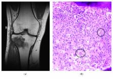
      Going Bone Deep: Osseous Rosai–Dorfman Disease in an Adult with Recurrent, Culture-Negative Osteomyelitis

      case-report

      Read this article at

      Bookmark
          There is no author summary for this article yet. Authors can add summaries to their articles on ScienceOpen to make them more accessible to a non-specialist audience.

          Abstract

          A patient presented for medical care on three separate occasions over the course of two years with recurrent right knee pain attributed to chronic osteomyelitis. Careful assessment revealed that his symptoms were caused by osseous Rosai–Dorfman disease. This case presents an alternative diagnostic possibility for culture-negative chronic osteomyelitis.

          Related collections

          Most cited references8

          • Record: found
          • Abstract: found
          • Article: not found

          Sinus histiocytosis with massive lymphadenopathy (Rosai-Dorfman disease): review of the entity.

          This report reviews the clinical and pathologic features of 423 examples of sinus histiocytosis with massive lymphadenopathy (SHML) entered in a case registry, with special emphasis on extranodal manifestations of the disease. The most common sites of extranodal involvement in this population are skin, upper respiratory tract, and bone. However, SHML also can occur in a variety of other sites, including the genitourinary system, lower respiratory tract, oral cavity, and soft tissues. Involvement of kidney, lower respiratory tract, or liver was found to be a poor prognostic sign, and patients with associated immunologic disease often fared poorly. In general, prognosis has been found to correlate both with the number of nodal groups and with the number of extranodal systems involved by SHML. A complete bibliography of publications describing patients with SHML is included, and illustrations of the clinical, histopathologic, and radiographic features of the disease are provided.
            Bookmark
            • Record: found
            • Abstract: found
            • Article: not found

            Langerhans' cell histiocytosis: pathology, imaging and treatment of skeletal involvement.

            Langerhans' cell histiocytosis (LCH) is manifested in a variety of ways, the most common being the eosinophilic granuloma, a localized, often solitary bone lesion that occurs predominantly in the pediatric age group. The hallmark of LCH is the proliferation and accumulation of a specific histiocyte: the Langerhans' cell. In bone this may cause pain and adjacent soft-tissue swelling, but some lesions are asymptomatic. LCH can involve any bone, but most lesions occur in the skull (especially the calvarium and temporal bones), the pelvis, spine, mandible, ribs, and tubular bones. Imaging diagnosis of the disease in bone is first based on the plain radiographic appearance, which is usually a central destructive, aggressive-looking lesion. In the skull, the lesions develop in the diploic space, are lytic, and their edges may be beveled, scalloped or confluent (geographic), or show a "button sequestrum." Vertebral body involvement usually causes collapse, resulting in vertebra plana. With significant recent improvements in the quality of gamma cameras, imaging techniques, and in studying children, bone scintigraphy at diagnosis and on follow-up usually reveals the sites of active disease, especially when the involvement is polyostotic. CT and MR imaging are very useful in providing detailed cross-sectional anatomic detail of the involved bone, including the bone marrow and the adjacent soft tissues. CT is better suited for demonstrating bone detail and MR imaging for bone marrow and soft-tissue involvement.
              Bookmark
              • Record: found
              • Abstract: not found
              • Article: not found

              Treatment of sinus histiocytosis with massive lymphadenopathy (rosai-dorfman disease): Report of a case and literature review

                Bookmark

                Author and article information

                Contributors
                Journal
                Case Rep Infect Dis
                Case Rep Infect Dis
                CRIID
                Case Reports in Infectious Diseases
                Hindawi
                2090-6625
                2090-6633
                2018
                17 May 2018
                : 2018
                : 6151738
                Affiliations
                1School of Medicine, University of North Carolina at Chapel Hill, Chapel Hill, NC, USA
                2Department of Medicine, Division of Infectious Diseases, School of Medicine, University of North Carolina at Chapel Hill, Chapel Hill, NC, USA
                3Department of Pathology and Laboratory Medicine, School of Medicine, University of North Carolina at Chapel Hill, Chapel Hill, NC, USA
                Author notes

                Academic Editor: Tomoyuki Shibata

                Author information
                http://orcid.org/0000-0002-4465-9830
                Article
                10.1155/2018/6151738
                5985093
                32374a74-410c-48c4-b74e-c160d092b7e4
                Copyright © 2018 Dominick S. DeFelice et al.

                This is an open access article distributed under the Creative Commons Attribution License, which permits unrestricted use, distribution, and reproduction in any medium, provided the original work is properly cited.

                History
                : 7 February 2018
                : 3 May 2018
                Funding
                Funded by: Doris Duke Charitable Foundation
                Categories
                Case Report

                Infectious disease & Microbiology
                Infectious disease & Microbiology

                Comments

                Comment on this article