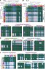
Summary Neuromodulatory control by oxytocin is essential to a wide range of social 1,2 , parental 3 and stress-related behaviors 4 . Autism spectrum disorders (ASD) are associated with deficiencies in oxytocin levels 5 and with genetic alterations of the oxytocin receptor (OXTR) 6 . Thirty years ago, Muhlethaler et al. 7 found that oxytocin increases the firing of inhibitory hippocampal neurons, but it remains unclear how elevated inhibition could account for the ability of oxytocin to improve information processing in the brain. Here, we describe a simple yet powerful mechanism by which oxytocin enhances cortical information transfer 8 while simultaneously lowering background activity, thus greatly improving signal-to-noise. Increased fast-spiking interneuron (FSI) activity not only suppresses spontaneous pyramidal cell firing, but also enhances the fidelity of spike transmission and sharpens spike timing. Use-dependent depression at the FSI-pyramidal cell synapse is both necessary and sufficient for the enhanced spike throughput. Notably, we show the generality of this novel circuit mechanism by activation of FSIs with cholecystokinin, or with channelrhodopsin-2. This provides insight into how a diffusely delivered neuromodulator can improve the performance of neural circuitry that requires synapse specificity and millisecond precision. Results The CA1 region of hippocampus receives potent excitatory input from neighboring area CA3 through the Schaffer Collateral (SC) pathway. Activation of SC axons evokes a monosynaptic excitatory post-synaptic potential (EPSP) onto CA1 pyramidal cells, as well as exciting a variety of CA1 interneurons. These interneurons then deliver a millisecond-delayed inhibitory postsynaptic potential (IPSP), termed feed-forward inhibition. Thus, both the stimulation threshold and the timing of spikes evoked in CA1 pyramidal cells by SC activation are dictated by a finely tuned balance of monosynaptic excitatory and disynaptic inhibitory inputs 8,9 . In agreement with previous results 8 , we found that stimulation of the SC pathway in acute rat hippocampal slices evoked spikes with a short latency and moderate jitter (Fig 1a). Strikingly, bath application of TGOT (Thr4,Gly7-Oxytocin, 200 nM), a specific agonist for oxytocin receptors, dramatically increased the probability of evoking a spike in the postsynaptic neuron from 0.50 to 0.82, while simultaneously suppressing the spontaneous activity of CA1 pyramidal cells by 57% from 1.4 Hz to 0.6 Hz (Fig 1a–d). The combination of increased evoked spike probability (signal) and reduced spontaneous activity (noise) resulted in an enhanced signal-to-noise. TGOT also reduced the latency and increased the temporal precision of evoked spikes (Fig 1e,f). In agreement with previous work 7,10 , TGOT increased the rate and amplitude of spontaneous inhibitory postsynaptic currents (IPSCs) onto CA1 pyramidal cells (Fig 1g, S1). Blockade by 10 µM bicuculline or by 100 nM tetrodotoxin indicated that these events were mediated by GABAA receptors and likely required an increase in interneuron firing rather than a change in spontaneous presynaptic release. The specific oxytocin receptor antagonist OTA ((d(CH2)5 1,Tyr(Me)2,Thr4,Orn8,des-Gly-NH2 9)-Vasotocin, 1 µM) blocked the TGOT-induced effects, suggesting that these actions were solely mediated by the oxytocin receptor 10 . The TGOT-induced increase in spontaneous IPSCs was also abolished by the potent P/Q-type calcium channel blocker ω-Agatoxin IVA, but unaffected by the N-type calcium channel antagonist ω-Conotoxin GVIA, suggesting that these events primarily arise from FSIs with little contribution from RS interneurons (Fig 1g, S1) 11,12 . To test more directly whether TGOT precisely targeted FS interneuron subtypes, we used whole cell recordings in CA1 strata oriens and pyramidale, stratum radiatum interneurons being unresponsive to TGOT 10 and lacking OXTR expression 13 . We found a clear distinction: FSIs were responsive to TGOT, whereas RS interneurons were not (Fig 2a). FSIs displayed robust responses upon application of 20 and 200 nM TGOT (Fig S2a–c), the latter producing a near-saturated effect. Dividing the increase in IPSCs onto pyramidal cells (27.3 Hz, Fig 1g) by the increase in FSI firing rate (8.8 Hz per FSI, Fig 2a), we calculate that on average each pyramidal cell receives input from at least ~3.1 TGOT-responsive FSIs in our slices. To clarify mechanisms by which TGOT depolarizes FSIs, we voltage clamped FS perisomatic-targeting (basket and axo-axonic) and RS basket cells at −65 mV. TGOT induced a large inward current in FSIs (Fig 2b, S2g), but as expected had no effect on the RS cells (data not shown). TGOT also increased the rate of spontaneous IPSCs onto the FSI (Fig S2d–f), as predicted from the FSI-FSI connectivity that may serve to regulate the distribution and extent of inhibition. To test whether the TGOT-induced inward current arises from G-protein signaling within the FSI itself, we replaced the GTP in the intracellular recording solution with 1 mM GTPγS, a non-hydrolysable GTP analog that renders G-proteins constitutively active. The GTPγS-induced current largely occluded the TGOT-induced current (Fig 2b, S2g), verifying that the TGOT effects involve G-protein signaling within the recorded neuron. The amplitude and kinetics of the TGOT-induced current were unaffected by intracellular BAPTA, indicating that the intracellular signaling mechanism is likely not Ca2+-dependent 14 . In voltage ramp recordings from FSIs, the TGOT-induced current reversed at −3.1±3.4 mV (Fig 2c, S2h), suggesting that the currents were generated by a non-selective cation channel. Partial replacement of external sodium by NMDG (50 mM) shifted the reversal potential to more negative values (−13.8±3.7 mV, P 2 min, at which point TGOT was applied. In 3 out of 13 recordings the voltage ramp-activated current (1) became more negative at all potentials shortly after TGOT application, and (2) failed to return to baseline after washout of the drug. It was assumed that this global shift was caused by a change in the space clamp or access resistance and these recordings were excluded from further analysis. Drugs and reagents All salts and buffers for intracellular and extracellular solutions, as well as ATP, GTP, GTPγS, phosphocreatine and biocytin were purchased from Sigma (St. Louis, MO). TGOT ((Thr4,Gly7)-Oxytocin), OTA ((d(CH2)5 1,Tyr(Me)2,Thr4,Orn8, des-Gly-NH2 9)-Vasotocin) and CCK (cholecystokinin octapeptide) peptides were purchased from Bachem (Torrance, CA), dissolved at 1 mM in ddH2O and stored at −20°C until use within 6 months of purchase. Bicuculline, TTX, NBQX, and D-AP5 were purchased from Ascent Scientific (Princeton, NJ). ω-conotoxin GVIA and ω-agatoxin IVA were purchased from Peptides International (Louisville, KY). Stock solutions were prepared and stored according to manufacturer specifications. Interneuron labeling and classification Physiological classification of interneuron subtypes was based on established criteria 11,25,31 . Fast-spiking cells were defined as those including (1) peak firing rates >200 Hz with little firing rate accommodation, (2) characteristic FS action potential waveform, and (3) minimal hyperpolarization-induced sag current due to Ih. Following interneuron recordings, slices were transferred to a fixative solution containing 4% paraformaldehyde, 0.2 % picric acid and 1× phosphate buffered saline for 24–72 h before being stained with 3,3’-diaminobenzidine tetrahydrochloride (0.015%) using a standard ABC kit (Vector). Neuronal cell types were identified based on morphology of axonal and dendritic arbors and electrophysiological properties of the cell. The FS perisomatic-targeting set includes both basket cells (shown), and axo-axonic cells (not shown). Because of technical challenges of discriminating FS basket and axo-axonic cells unequivocally, both cell types were pooled into a single group of FS perisomatic-targeting cells. When analyzed separately, both putative types were equivalently responsive to TGOT. Analysis of cell-attached and intracellular recording data Analysis of spikes, evoked synaptic currents, and synaptic potentials were performed offline using custom written routines in MATLAB (Mathworks). Spontaneous IPSCs were detected using a modified version of the detectPSPs script by Phil Larimer (http://www.mathworks.com/matlabcentral/fileexchange). Spike jitter histograms were calculated by subtracting the latency of each spike from the average latency of spikes evoked in that cell. The average latency and jitter were calculated separately for control and TGOT/CCK conditions in each cell. To measure the spike width, raw data was oversampled to 133 kHz using the MATLAB spline function. Time course of spontaneous activity in pyramidal cell attached recordings was calculated by averaging over all cells and smoothing in time with a boxcar filter (width=7 sweeps). Optical stimulation of channelrhodopsin-2 Photostimuli were produced by three Luxeon Rebel LEDs (470 nm, Philips Lumileds, San Jose, CA) driven by a custom-built controller. The LEDs were placed below the recording chamber for full slice illumination once stable recording conditions were reached. Light pulses were 5 ms in duration with a power of approximately 0.5 mW/mm2. ChR2-evoked IPSCs were recorded from CA1 pyramidal neurons (n=10 cells, N=4 animals). Six of these neurons were recorded in the same region of the same slice as neurons recorded in the cell-attached data set. Data analysis for cell-attached recordings involving blue light stimulation In the full data set, blue light stimulation increased the spike probability in 13 of 16 neurons (Fig S8a, 12% increase in spike probability including all neurons, P<0.05, two-tailed t-test). In recordings from rat neurons the average increase in spike firing probability with TGOT or CCK was not correlated with spike latency, whereas in the mouse data we found a strong correlation between control spike latency and the ChR2-induced increase in spike probability (Fig S8d,e). In the mouse data set, the shortest latency spikes showed the weakest increase in spike firing probability. Plotting the latency against the jitter of spikes elicited under control conditions, we found a clear separation between two groups of cells, in which evoked spikes from one set of cells occurred with very short latency and little jitter and spikes from another set of cells occurred at longer latency and with more jitter (Fig S8f). Because of the smaller size of the mouse brain, we found our slice angle to be less reliably transverse than in the rat preparation. As a result, the stimulating and recording electrodes were placed closer to one another in the mouse slice in order for the stimulating electrode to recruit a sufficient number of excitatory Schaffer Collateral fibers to drive an action potential in the postsynaptic CA1 pyramidal cell. This change in recording configuration unfortunately increases the probability of directly activating inhibitory fibers with the stimulating electrode and generating a monosynaptic IPSC. A well-documented set of physiological parameters, including synaptic kinetics and cell excitability 25 ensure that the physiologically relevant disynaptic IPSC arises mostly from FSIs. The monosynaptically activated IPSC, however, will arise from a less targeted subset of neurons, and therefore be less susceptible to modulation by interventions that selectively target FSIs. In the cell-attached recording configuration, it was impossible to determine directly the relative monosynaptic and disynaptic contributions to the feed-forward IPSC. The monosynaptic IPSC relies only on a single GABAergic synapse, however, whereas the disynaptic IPSC relies on three sequential steps: (1) a glutamatergic synapse onto the interneuron, (2) the subsequent action potential in the interneuron, and finally (3) the GABAergic transmission onto the postsynaptic pyramidal cell. The monosynaptic IPSC will therefore occur with a shorter latency and less jitter than the disynaptically evoked IPSC. As a result, spikes in pyramidal cells in which the feed-forward IPSC is dominated by a monosynaptic component will be expected to occur with a shorter latency and less jitter than spikes in cells experiencing a more physiological disynaptic feed-forward IPSC. We therefore excluded the tightly clustered group of neurons with very short latency and low jitter spikes from the mouse data set (N=9 cells) and analyzed only the neurons in which spikes occurred with a longer latency and more jitter, consistent with disynaptic feed-forward inhibition (N=7 cells). All of these remaining cells demonstrated an increase in spike firing probability following blue light stimulation (7 out of 7 cells, 28% increase in spike probability; P<0.01 paired two-tailed t-test). In the complete data set (N = 16 cells) we observed a modest increase in spike latency following blue light stimulation of PV interneurons across all 16 neurons (Fig S8b,c). However, in the 5 out of 7 cells from the restricted data set that fired at least 5 spikes in both the control and blue light stimulation conditions, light activation of PV interneurons reduced the latency (Fig S8g, 10.35 ms in control, 10.07 ms following light stimulation; P = 0.73 paired two-tailed t-test) and jitter (Fig S8h, 16.58 ms2 control; 11.78 ms2 light stimulation; P = 0.23 paired two-tailed t-test) of spikes. Although this reduction in latency and jitter did not reach statistical significance, the trend is consistent with our TGOT and CCK results. Immunohistochemistry At the end of each ChR2 recording session, slices were fixed overnight with 4% paraformaldehyde (PFA)/phosphate buffered saline (PBS) solution and cryoprotected by immersion in 30% sucrose/PBS solution overnight at 4°C. Tissues were embedded in Tissue Tek, frozen on dry ice, and cryosectioned at 20 µm thickness. Sections for were processed using 1.5% normal goat serum (NGS) and 0.1% Triton X-100 in all procedures except washing steps, where only PBS was used. Sections were incubated in blocking solution for 1 hr, followed by incubation with the primary antibodies overnight at 4°C. Cryostat tissue sections were stained with the primary antibodies: mouse anti-Parvalbumin (1:1000, Sigma) and rabbit anti-DsRed (1:500, Chemicon). Secondary antibodies conjugated with Alexa fluoro dyes 488, 594 (Molecular Probes) raised from the same host used for blocking serum were applied for 1 hr at room temperature. Nuclear counterstaining was performed with 100 ng/ml 4,6-diamidino-2-phenylindole (DAPI) solution in PBS for 5 min. Fluorescent images were captured using a cooled-CCD camera (Princeton Scientific Instruments, NJ) using Metamorph software (Universal imaging, Downingtown, Pennsylvania). Virus injection Adeno-associated virus carrying ChR2 fused to the fluorescent marker mCherry AAV2/1.EF1.dflox.hChR2(H134R)-mCherry.WPRE.hGH, (University of Pennsylvania Gene Therapy Program Vector Core) was injected bilaterally into dorsal hippocampal CA1 region of Pvalb-cre (PV-Cre) transgenic mice 32 (aged between postnatal days 15–19) at three sites: 2.2, 1.8 and 1.6 mm posterior from bregma, 2.4, 2.1, 1.7 mm from midline, and 1.2, 1.1, and 1 mm below cortical surface, respectively. Animals were anesthetized with isoflurane, mounted in a stereotactic apparatus and kept under isoflurane anesthesia during surgery. We injected 100 nL of virus at each location over a 2 min period using a glass micropipette (tip diameter ~20 µm) attached to a Nanoliter 2000 pressure injection apparatus (World Precision Instruments). The pipette was held in place for 3 min following each injection before being completely retracted from the brain. Mice were returned to their home cage for 2–3 weeks before acute slice preparation to allow for virus expression. Computational model of EPSP-spike coupling The computater modeling was performed using NEURON and automated using MATLAB. A simplified pyramidal cell, consisting of a soma, a single axon and a single dendrite was initialized to starting parameters before each stimulus. Background and voltage-gated conductances were based on reported models 33,34 . Small adjustments were made to improve agreement of parameters such as cell excitability and action potential waveform between the model and experimental observations. Each sweep consisted of (1) a “monosynaptic” EPSC onto the dendrite, (2) a “disyanptic” feed-forward IPSC onto the soma and dendrite 2 ms after the evoked EPSC (unless otherwise specified), and (3) multiple “spontaneous” IPSCs onto the soma with randomly distributed amplitudes and timing. To isolate the role of the feed-forward IPSC from changes in inhibitory-tone, spontaneous IPSCs were omitted in the simulation used to generate Fig 4h,i. At the outset of each set of sweeps, the “evoked” EPSC-IPSC amplitudes were set empirically by increasing the EPSC and IPSC conductances together with a fixed ratio of 6:1 until ~50% chance of spike propagation was reached. Experimental measurement of IPSC/EPSC ratio ranged from 2.62 to 5.20 (mean±S.E.M. of 3.65±0.28). This experimentally measured range is presumed to be an underestimate of the true ratio due to imperfect isolation of the IPSC reversal potential, causing a presumed GABAergic contribution to the measured EPSC in some cells. In the model, IPSC/EPSC ratios from 4:1 up to 6:1 showed a pronounced TGOT-induced increase in evoked spike probability, with 6:1 supporting the strongest influence of TGOT on spike timing and jitter. Variability was introduced by using pseudo-random number generation to vary independently (1) the evoked EPSC conductance, (2) the evoked IPSC conductance and (3) the spontaneous IPSC timing and amplitudes. Evoked EPSC and IPSC conductances were varied independently on each sweep according to a normal distribution centered on the empirically determined mean value, with a standard deviation that was 5% of the mean. TGOT was simulated by (1) reducing the evoked somatic IPSC conductance to 60% of “baseline”, while sparing the evoked EPSC and the dendritic IPSC, (2) doubling the spontaneous IPSC amplitude, and (3) increasing the spontaneous IPSC rate from 5 Hz to 35 Hz. The IPSC reversal potential was set at −110 mV for Fig S10b–c, consistent with the calculated GABAA reversal potential in our whole cell recording conditions. For the rest of the simulations, the IPSC reversal potential was set to −90 mV, consistent with cell-attached recording conditions. The increase in evoked spike probability was robust as the GABAA reversal potential was varied from −80 mV to −120 mV (Fig S12), while the reduction in latency and latency jitter were decreased in magnitude but remained statistically significant. Supplementary Material 1