1
views
0
recommends
+1 Recommend
0 collections
    0
    shares
      • Record: found
      • Abstract: found
      • Article: found
      Is Open Access

      Primary intradural extramedullary Ewing sarcoma: A case report and literature review

      research-article

      Read this article at

      Bookmark
          There is no author summary for this article yet. Authors can add summaries to their articles on ScienceOpen to make them more accessible to a non-specialist audience.

          Abstract

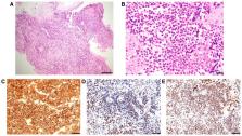
          Tumors of the spinal cord and cauda equina show a wide spectrum of histology and require careful diagnosis and treatment. Primary intradural extramedullary Ewing sarcoma (IEES) is extremely rare, and initial imaging and clinical findings for this tumor mimic those of benign intradural spinal tumors. The present report describes a case of a 35-year-old woman who presented with IEES with meningeal seeding, and the literature on the management and clinical course of these tumors was reviewed. An examination revealed right-side leg dominant rapidly progressive hypoesthesia and motor deficits. MRI identified intradural tumors at the T12-L1 and L4-5 levels. Growth and an increase in the number of intradural tumors occurred over 3 weeks. Gadolinium-contrast T1-weighted MRI revealed diffusely enhanced signals for tumorous lesions with meningeal seeding in the cervical and thoracic spinal cord. Excision of a tumor at T12-L1 was performed to alleviate the symptoms and to make a histologic diagnosis. Microscopically, the tumor consisted of dense sheets of small round cells. Immunohistochemically, tumor cells showed intense and diffuse positive staining for CD99, ETS transcription factor ERG and Fli-1 proto-oncogene, ETS transcription factor (FLI1). The sequence analysis revealed the EWS RNA binding protein 1-FLI1 fusion transcript. The lesion was diagnosed as primary intradural ES. Adjuvant chemotherapy following radiotherapy for the whole spine was performed; however, multiple brain metastases were found at 10 months after diagnosis and the patient died of diffusely disseminated disease limited to the central nervous system without evidence of distant metastases at 16 months after the initial diagnosis. In a literature review of IEES cases, the 1- and 5-year overall survival rates were 79.8 and 26.6%, respectively, and the 1-, 2- and 5-year progression-free survival rates were 61.0, 52.3 and 10.9%, respectively. Therefore, primary IEES has a poor prognosis compared with ES of bone, and novel agents and treatment strategies are required.

          Related collections

          Most cited references32

          • Record: found
          • Abstract: found
          • Article: not found

          Ewing Sarcoma: Current Management and Future Approaches Through Collaboration.

          Ewing sarcoma (ES) is an aggressive sarcoma of bone and soft tissue occurring at any age with a peak incidence in adolescents and young adults. The treatment of ES relies on a multidisciplinary approach, coupling risk-adapted intensive neoadjuvant and adjuvant chemotherapies with surgery and/or radiotherapy for control of the primary site and possible metastatic disease. The optimization of ES multimodality therapeutic strategies has resulted from the efforts of several national and international groups in Europe and North America and from cooperation between pediatric and medical oncologists. Successive first-line trials addressed the efficacy of various cyclic combinations of drugs incorporating doxorubicin, vincristine, cyclophosphamide, ifosfamide, etoposide, and dactinomycin and identified prognostic factors now used to tailor therapies. The role of high-dose chemotherapy is still debated. Current 5-year overall survival for patients with localized disease is 65% to 75%. Patients with metastases have a 5-year overall survival < 30%, except for those with isolated pulmonary metastasis (approximately 50%). Patients with recurrence have a dismal prognosis. The many insights into the biology of the EWS-FLI1 protein in the initiation and progression of ES remain to be translated into novel therapeutic strategies. Current options and future approaches will be discussed.
            Bookmark
            • Record: found
            • Abstract: found
            • Article: not found

            Clinical features and outcomes in patients with extraskeletal Ewing sarcoma.

            Ewing sarcoma can arise in either bone or soft tissue. The purpose of this study was to investigate whether patient characteristics, treatment strategies, and outcomes differ between skeletal Ewing sarcoma and extraskeletal Ewing sarcoma (EES). Patients <40 years of age with Ewing sarcoma or peripheral primitive neuroectodermal tumor reported to the United States Surveillance, Epidemiology, and End Results Program database from 1973 to 2007 were evaluated based on skeletal (n = 1519) versus extraskeletal (n = 683) site of origin. Patient characteristics were compared using Fisher exact tests. Overall survival was estimated via the Kaplan-Meier method and compared using log-rank tests and Cox proportional hazard models. Patients with EES had a higher mean age (19.5 vs 16.3 years; P < .001) and were less likely to be male (53.4% vs 63.3%; P < .001) or white (84.8% vs 92.5%; P < .001) compared with patients with skeletal tumors. Extraskeletal tumors were more likely to arise in axial locations (72.9% vs 54.2%; P = .001) but were less likely to arise specifically in the pelvis (19.8% vs 26.6%; P < .001). Metastatic status or tumor size did not differ by group. Five-year overall survival was superior for localized EES compared with localized skeletal tumors (69.7% vs 62.6%; P = .02). The hazard ratio for death in patients with localized skeletal tumors compared with localized EES was 2.36 (95% confidence interval, 1.61-3.44) beyond 24 months from initial diagnosis. Patient characteristics and outcomes differ among patients with EES compared with patients with skeletal Ewing sarcoma. These findings may have important implications for patient care. Copyright © 2011 American Cancer Society.
              Bookmark
              • Record: found
              • Abstract: found
              • Article: not found

              Spinal tumors.

              Spinal tumors are uncommon lesions but may cause significant morbidity in terms of limb dysfunction. In establishing the differential diagnosis for a spinal lesion, location is the most important feature, but the clinical presentation and the patient's age and gender are also important. Magnetic resonance (MR) imaging plays a central role in the imaging of spinal tumors, easily allowing tumors to be classified as extradural, intradural-extramedullary or intramedullary, which is very useful in tumor characterization. In the evaluation of lesions of the osseous spine both computed tomography (CT) and MR are important. We describe the most common spinal tumors in detail. In general, extradural lesions are the most common with metastasis being the most frequent. Intradural tumors are rare, and the majority is extramedullary, with meningiomas and nerve sheath tumors being the most frequent. Intramedullary tumors are uncommon spinal tumors. Astrocytomas and ependymomas comprise the majority of the intramedullary tumors. The most important tumors are documented with appropriate high quality CT or MR images and the characteristics of these tumors are also summarized in a comprehensive table. Finally we illustrate the use of the new World Health Organization (WHO) classification of neoplasms affecting the central nervous system.
                Bookmark

                Author and article information

                Journal
                Oncol Lett
                Oncol Lett
                OL
                Oncology Letters
                D.A. Spandidos
                1792-1074
                1792-1082
                September 2020
                26 June 2020
                26 June 2020
                : 20
                : 3
                : 2347-2355
                Affiliations
                [1 ]Department of Orthopaedics and Rehabilitation Medicine, University of Fukui Faculty of Medical Sciences, Eiheiji, Fukui 910-1193, Japan
                [2 ]Division of Diagnostic Pathology/Surgical Pathology, University of Fukui Hospital, Eiheiji, Fukui 910-1193, Japan
                [3 ]Department of Pathology, Kanazawa University, Kanazawa, Ishikawa 920-8641, Japan
                Author notes
                Correspondence to: Dr Hideaki Nakajima, Department of Orthopaedics and Rehabilitation Medicine, University of Fukui Faculty of Medical Sciences, 23-3 Matsuoka Shimoaizuki, Eiheiji, Fukui 910-1193, Japan, E-mail: nhideaki@ 123456u-fukui.ac.jp
                Article
                OL-0-0-11786
                10.3892/ol.2020.11786
                7401003
                32782552
                31f12212-e8d8-4be0-b016-9feb476df275
                Copyright: © Izubuchi et al.

                This is an open access article distributed under the terms of the Creative Commons Attribution-NonCommercial-NoDerivs License, which permits use and distribution in any medium, provided the original work is properly cited, the use is non-commercial and no modifications or adaptations are made.

                History
                : 21 January 2020
                : 27 May 2020
                Categories
                Articles

                Oncology & Radiotherapy
                ewing sarcoma,intradural tumor,cauda equina tumor,radiotherapy,adjuvant chemotherapy,meningeal seeding

                Comments

                Comment on this article