- Record: found
- Abstract: found
- Article: found
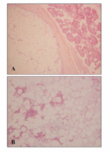
A case of lipomatous pleomorphic adenoma in the parotid gland: a case report
research-article
Read this article at
There is no author summary for this article yet. Authors can add summaries to their articles on ScienceOpen to make them more accessible to a non-specialist audience.
Abstract
Introduction
Pleomorphic adenoma is the most common benign neoplasm of the salivary glands. Extensive lipomatous involvement of the tumor is, however, a very rare finding.
Related collections
Most cited references5
- Record: found
- Abstract: found
- Article: not found
Lipomatous pleomorphic adenoma of the parotid gland. Classification of lipomatous tissue in salivary glands.
G. Seifert, K Donath, R. Schäfer (1999)
- Record: found
- Abstract: found
- Article: not found
Myxolipomatous pleomorphic adenoma: an unusual oral presentation.
Fumio Ide, Kaoru Kusama (2003)
- Record: found
- Abstract: not found
- Article: not found
Pleomorphic adenoma with extensive lipometaplasia.
L. Ma, Enders K W Ng (1995)