25
views
0
recommends
+1 Recommend
0 collections
    0
    shares
      • Record: found
      • Abstract: found
      • Article: not found

      Clostridium perfringens Enterotoxin Fragment Removes Specific Claudins from Tight Junction Strands : Evidence for Direct Involvement of Claudins in Tight Junction Barrier

      research-article

      Read this article at

      ScienceOpenPublisherPMC
      Bookmark
          There is no author summary for this article yet. Authors can add summaries to their articles on ScienceOpen to make them more accessible to a non-specialist audience.

          Abstract

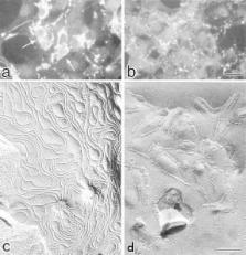
          Claudins, comprising a multigene family, constitute tight junction (TJ) strands. Clostridium perfringens enterotoxin (CPE), a single ∼35-kD polypeptide, was reported to specifically bind to claudin-3/RVP1 and claudin-4/CPE-R at its COOH-terminal half. We examined the effects of the COOH-terminal half fragment of CPE (C-CPE) on TJs in L transfectants expressing claudin-1 to -4 (C1L to C4L, respectively), and in MDCK I cells expressing claudin-1 and -4. C-CPE bound to claudin-3 and -4 with high affinity, but not to claudin-1 or -2. In the presence of C-CPE, reconstituted TJ strands in C3L cells gradually disintegrated and disappeared from their cell surface. In MDCK I cells incubated with C-CPE, claudin-4 was selectively removed from TJs with its concomitant degradation. At 4 h after incubation with C-CPE, TJ strands were disintegrated, and the number of TJ strands and the complexity of their network were markedly decreased. In good agreement with the time course of these morphological changes, the TJ barrier (TER and paracellular flux) of MDCK I cells was downregulated by C-CPE in a dose-dependent manner. These findings provided evidence for the direct involvement of claudins in the barrier functions of TJs.

          Related collections

          Most cited references36

          • Record: found
          • Abstract: found
          • Article: not found

          Claudin-1 and -2: Novel Integral Membrane Proteins Localizing at Tight Junctions with No Sequence Similarity to Occludin

          Occludin is the only known integral membrane protein localizing at tight junctions (TJ), but recent targeted disruption analysis of the occludin gene indicated the existence of as yet unidentified integral membrane proteins in TJ. We therefore re-examined the isolated junction fraction from chicken liver, from which occludin was first identified. Among numerous components of this fraction, only a broad silver-stained band ∼22 kD was detected with the occludin band through 4 M guanidine-HCl extraction as well as sonication followed by stepwise sucrose density gradient centrifugation. Two distinct peptide sequences were obtained from the lower and upper halves of the broad band, and similarity searches of databases allowed us to isolate two full-length cDNAs encoding related mouse 22-kD proteins consisting of 211 and 230 amino acids, respectively. Hydrophilicity analysis suggested that both bore four transmembrane domains, although they did not show any sequence similarity to occludin. Immunofluorescence and immunoelectron microscopy revealed that both proteins tagged with FLAG or GFP were targeted to and incorporated into the TJ strand itself. We designated them as “claudin-1” and “claudin-2”, respectively. Although the precise structure/function relationship of the claudins to TJ still remains elusive, these findings indicated that multiple integral membrane proteins with four putative transmembrane domains, occludin and claudins, constitute TJ strands.
            Bookmark
            • Record: found
            • Abstract: found
            • Article: not found

            A Single Gene Product, Claudin-1 or -2, Reconstitutes Tight Junction Strands and Recruits Occludin in Fibroblasts

            Three integral membrane proteins, clau- din-1, -2, and occludin, are known to be components of tight junction (TJ) strands. To examine their ability to form TJ strands, their cDNAs were introduced into mouse L fibroblasts lacking TJs. Immunofluorescence microscopy revealed that both FLAG-tagged claudin-1 and -2 were highly concentrated at cell contact sites as planes through a homophilic interaction. In freeze-fracture replicas of these contact sites, well-developed networks of strands were identified that were similar to TJ strand networks in situ and were specifically labeled with anti-FLAG mAb. In glutaraldehyde-fixed samples, claudin-1–induced strands were largely associated with the protoplasmic (P) face as mostly continuous structures, whereas claudin-2–induced strands were discontinuous at the P face with complementary grooves at the extracellular (E) face which were occupied by chains of particles. Although occludin was also concentrated at cell contact sites as dots through its homophilic interaction, freeze-fracture replicas identified only a small number of short strands that were labeled with anti-occludin mAb. However, when occludin was cotransfected with claudin-1, it was concentrated at cell contact sites as planes to be incorporated into well- developed claudin-1–based strands. These findings suggested that claudin-1 and -2 are mainly responsible for TJ strand formation, and that occludin is an accessory protein in some function of TJ strands.
              Bookmark
              • Record: found
              • Abstract: found
              • Article: not found

              Functional dissociation of paracellular permeability and transepithelial electrical resistance and disruption of the apical- basolateral intramembrane diffusion barrier by expression of a mutant tight junction membrane protein

              Tight junctions, the most apical of the intercellular junctions that connect individual cells in a epithelial sheet, are thought to form a seal that restricts paracellular and intramembrane diffusion. To analyze the functioning of tight junctions, we generated stable MDCK strain 2 cell lines expressing either full-length or COOH-terminally truncated chicken occludin, the only known transmembrane component of tight junctions. Confocal immunofluorescence and immunoelectron microscopy demonstrated that mutant occludin was incorporated into tight junctions but, in contrast to full-length chicken occludin, exhibited a discontinuous junctional staining pattern and also disrupted the continuous junctional ring formed by endogenous occludin. This rearrangement of occludin was not paralleled by apparent changes in the junctional morphology as seen by thin section electron microscopy nor apparent discontinuities of the junctional strands observed by freeze-fracture. Nevertheless, expression of both wild-type and mutant occludin induced increased transepithelial electrical resistance (TER). In contrast to TER, particularly the expression of COOH-terminally truncated occludin led to a severalfold increase in paracellular flux of small molecular weight tracers. Since the selectivity for size or different types of cations was unchanged, expression of wild-type and mutant occludin appears to have activated an existing mechanism that allows selective paracellular flux in the presence of electrically sealed tight junctions. Occludin is also involved in the formation of the apical/basolateral intramembrane diffusion barrier, since expression of the COOH-terminally truncated occludin was found to render MDCK cells incapable of maintaining a fluorescent lipid in a specifically labeled cell surface domain.
                Bookmark

                Author and article information

                Contributors
                Journal
                J Cell Biol
                The Journal of Cell Biology
                The Rockefeller University Press
                0021-9525
                1540-8140
                4 October 1999
                : 147
                : 1
                : 195-204
                Affiliations
                [a ]Department of Cell Biology, Faculty of Medicine, Kyoto University, Sakyo-ku, Kyoto 606-8501, Japan
                [b ]Project Research for Molecular Bacteriology, Research Institute for Microbial Diseases, Osaka University 3-1 Yamadaoka, Suita, Osaka 565-0871, Japan
                [c ]Laboratory of Cell Biology, KAN Research Institute Inc., Kyoto Research Park, Chudoji, Shimogyo-ku, Kyoto 600-8317, Japan
                [d ]Department of Molecular Cell Biology, Institute of DNA Medicine, The Jikei University School of Medicine, Nishi-Shinbashi, Minato-ku, Tokyo 105-8461, Japan
                Article
                9907056
                10.1083/jcb.147.1.195
                2164970
                10508866
                2e954c9f-b7f3-45a1-bc64-83d68e72bd83
                © 1999 The Rockefeller University Press
                History
                : 14 July 1999
                : 17 August 1999
                : 31 August 1999
                Categories
                Original Article

                Cell biology
                clostridium perfringens enterotoxin,tight junction,claudin,epithelial barrier,freeze-fracture

                Comments

                Comment on this article