37
views
0
recommends
+1 Recommend
0 collections
    0
    shares
      • Record: found
      • Abstract: found
      • Article: found
      Is Open Access

      Questions and controversies: the role of necroptosis in liver disease

      review-article
      1 , 2 , * , 1 , 2 , 1 , 2
      Cell Death Discovery
      Nature Publishing Group

      Read this article at

      Bookmark
          There is no author summary for this article yet. Authors can add summaries to their articles on ScienceOpen to make them more accessible to a non-specialist audience.

          Abstract

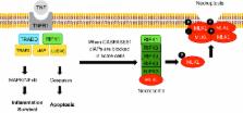
          Acute and chronic liver injury results in hepatocyte death and turnover. If injury becomes chronic, the continuous cell death and turnover leads to chronic inflammation, fibrosis and ultimately cirrhosis and hepatocellular carcinoma. Controlling liver cell death both in acute injury, to rescue the liver from acute liver failure, and in chronic injury, to curb secondary inflammation and fibrosis, is of paramount importance as a therapeutic strategy. Both apoptosis and necrosis occur in the liver, but the occurrence of necroptosis in the liver and its contribution to liver disease is controversial. Necroptosis is a form of regulated necrosis which occurs in certain cell types when caspases (+/−cIAPs) are inhibited through the RIPK1-RIPK3 activation of MLKL. The occurrence of necroptosis in the liver has recently been examined in multiple liver injury models with conflicting results. The aim of this review is to summarize the published data with an emphasis on the controversies and remaining questions in the field.

          Related collections

          Most cited references73

          • Record: found
          • Abstract: found
          • Article: not found

          Mouse model of chronic and binge ethanol feeding (the NIAAA model).

          Chronic alcohol consumption is a leading cause of chronic liver disease worldwide, leading to cirrhosis and hepatocellular carcinoma. Currently, the most widely used model for alcoholic liver injury is ad libitum feeding with the Lieber-DeCarli liquid diet containing ethanol for 4-6 weeks; however, this model, without the addition of a secondary insult, only induces mild steatosis, slight elevation of serum alanine transaminase (ALT) and little or no inflammation. Here we describe a simple mouse model of alcoholic liver injury by chronic ethanol feeding (10-d ad libitum oral feeding with the Lieber-DeCarli ethanol liquid diet) plus a single binge ethanol feeding. This protocol for chronic-plus-single-binge ethanol feeding synergistically induces liver injury, inflammation and fatty liver, which mimics acute-on-chronic alcoholic liver injury in patients. This feeding protocol can also be extended to chronic feeding for longer periods of time up to 8 weeks plus single or multiple binges. Chronic-binge ethanol feeding leads to high blood alcohol levels; thus, this simple model will be very useful for the study of alcoholic liver disease (ALD) and of other organs damaged by alcohol consumption.
            Bookmark
            • Record: found
            • Abstract: found
            • Article: found
            Is Open Access

            Translocation of mixed lineage kinase domain-like protein to plasma membrane leads to necrotic cell death

            Mixed lineage kinase domain-like protein (MLKL) was identified to function downstream of receptor interacting protein 3 (RIP3) in tumor necrosis factor-α (TNF)-induced necrosis (also called necroptosis). However, how MLKL functions to mediate necroptosis is unknown. By reconstitution of MLKL function in MLKL-knockout cells, we showed that the N-terminus of MLKL is required for its function in necroptosis. The oligomerization of MLKL in TNF-treated cells is essential for necroptosis, as artificially forcing MLKL together by using the hormone-binding domain (HBD*) triggers necroptosis. Notably, forcing together the N-terminal domain (ND) but not the C-terminal kinase domain of MLKL causes necroptosis. Further deletion analysis showed that the four-α-helix bundle of MLKL (1-130 amino acids) is sufficient to trigger necroptosis. Both the HBD*-mediated and TNF-induced complexes of MLKL(ND) or MLKL are tetramers, and translocation of these complexes to lipid rafts of the plasma membrane precedes cell death. The homo-oligomerization is required for MLKL translocation and the signal sequence for plasma membrane location is located in the junction of the first and second α-helices of MLKL. The plasma membrane translocation of MLKL or MLKL(ND) leads to sodium influx, and depletion of sodium from the cell culture medium inhibits necroptosis. All of the above phenomena were not seen in apoptosis. Thus, the MLKL oligomerization leads to translocation of MLKL to lipid rafts of plasma membrane, and the plasma membrane MLKL complex acts either by itself or via other proteins to increase the sodium influx, which increases osmotic pressure, eventually leading to membrane rupture.
              Bookmark
              • Record: found
              • Abstract: found
              • Article: not found

              Activation of the pseudokinase MLKL unleashes the four-helix bundle domain to induce membrane localization and necroptotic cell death.

              Necroptosis is considered to be complementary to the classical caspase-dependent programmed cell death pathway, apoptosis. The pseudokinase Mixed Lineage Kinase Domain-Like (MLKL) is an essential effector protein in the necroptotic cell death pathway downstream of the protein kinase Receptor Interacting Protein Kinase-3 (RIPK3). How MLKL causes cell death is unclear, however RIPK3-mediated phosphorylation of the activation loop in MLKL trips a molecular switch to induce necroptotic cell death. Here, we show that the MLKL pseudokinase domain acts as a latch to restrain the N-terminal four-helix bundle (4HB) domain and that unleashing this domain results in formation of a high-molecular-weight, membrane-localized complex and cell death. Using alanine-scanning mutagenesis, we identified two clusters of residues on opposing faces of the 4HB domain that were required for the 4HB domain to kill cells. The integrity of one cluster was essential for membrane localization, whereas MLKL mutations in the other cluster did not prevent membrane translocation but prevented killing; this demonstrates that membrane localization is necessary, but insufficient, to induce cell death. Finally, we identified a small molecule that binds the nucleotide binding site within the MLKL pseudokinase domain and retards MLKL translocation to membranes, thereby preventing necroptosis. This inhibitor provides a novel tool to investigate necroptosis and demonstrates the feasibility of using small molecules to target the nucleotide binding site of pseudokinases to modulate signal transduction.
                Bookmark

                Author and article information

                Journal
                Cell Death Discov
                Cell Death Discov
                Cell Death Discovery
                Nature Publishing Group
                2058-7716
                05 December 2016
                2016
                : 2
                : 16089
                Affiliations
                [1 ]Research Center for Liver Disease, Keck School of Medicine, Department of Medicine, University of Southern California , Los Angeles, CA, USA
                [2 ]Division of GI/Liver, Department of Medicine, Keck School of Medicine, University of Southern California , Los Angeles, CA, USA
                Author notes
                Article
                cddiscovery201689
                10.1038/cddiscovery.2016.89
                5136616
                27924226
                2dd7f439-a8f1-4c96-bbb1-a8fd77e0cda1
                Copyright © 2016 The Author(s)

                This work is licensed under a Creative Commons Attribution 4.0 International License. The images or other third party material in this article are included in the article’s Creative Commons license, unless indicated otherwise in the credit line; if the material is not included under the Creative Commons license, users will need to obtain permission from the license holder to reproduce the material. To view a copy of this license, visit http://creativecommons.org/licenses/by/4.0/

                History
                : 13 June 2016
                : 09 October 2016
                : 17 October 2016
                Categories
                Review Article

                Comments

                Comment on this article