4
views
0
recommends
+1 Recommend
0 collections
    0
    shares
      • Record: found
      • Abstract: found
      • Article: found
      Is Open Access

      Central encoding of the strength of intranasal chemosensory trigeminal stimuli in a human experimental pain setting

      research-article

      Read this article at

      Bookmark
          There is no author summary for this article yet. Authors can add summaries to their articles on ScienceOpen to make them more accessible to a non-specialist audience.

          Abstract

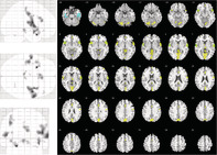
          An important measure in pain research is the intensity of nociceptive stimuli and their cortical representation. However, there is evidence of different cerebral representations of nociceptive stimuli, including the fact that cortical areas recruited during processing of intranasal nociceptive chemical stimuli included those outside the traditional trigeminal areas. Therefore, the aim of this study was to investigate the major cerebral representations of stimulus intensity associated with intranasal chemical trigeminal stimulation. Trigeminal stimulation was achieved with carbon dioxide presented to the nasal mucosa. Using a single‐blinded, randomized crossover design, 24 subjects received nociceptive stimuli with two different stimulation paradigms, depending on the just noticeable differences in the stimulus strengths applied. Stimulus‐related brain activations were recorded using functional magnetic resonance imaging with event‐related design. Brain activations increased significantly with increasing stimulus intensity, with the largest cluster at the right Rolandic operculum and a global maximum in a smaller cluster at the left lower frontal orbital lobe. Region of interest analyses additionally supported an activation pattern correlated with the stimulus intensity at the piriform cortex as an area of special interest with the trigeminal input. The results support the piriform cortex, in addition to the secondary somatosensory cortex, as a major area of interest for stimulus strength‐related brain activation in pain models using trigeminal stimuli. This makes both areas a primary objective to be observed in human experimental pain settings where trigeminal input is used to study effects of analgesics.

          Abstract

          Besides the secondary somatosensory cortex, the human piriform cortex is a major area of interest for stimulus strength‐related brain activation by trigeminal nociceptive stimuli.

          Related collections

          Most cited references77

          • Record: found
          • Abstract: found
          • Article: not found

          Automated anatomical labeling of activations in SPM using a macroscopic anatomical parcellation of the MNI MRI single-subject brain.

          An anatomical parcellation of the spatially normalized single-subject high-resolution T1 volume provided by the Montreal Neurological Institute (MNI) (D. L. Collins et al., 1998, Trans. Med. Imag. 17, 463-468) was performed. The MNI single-subject main sulci were first delineated and further used as landmarks for the 3D definition of 45 anatomical volumes of interest (AVOI) in each hemisphere. This procedure was performed using a dedicated software which allowed a 3D following of the sulci course on the edited brain. Regions of interest were then drawn manually with the same software every 2 mm on the axial slices of the high-resolution MNI single subject. The 90 AVOI were reconstructed and assigned a label. Using this parcellation method, three procedures to perform the automated anatomical labeling of functional studies are proposed: (1) labeling of an extremum defined by a set of coordinates, (2) percentage of voxels belonging to each of the AVOI intersected by a sphere centered by a set of coordinates, and (3) percentage of voxels belonging to each of the AVOI intersected by an activated cluster. An interface with the Statistical Parametric Mapping package (SPM, J. Ashburner and K. J. Friston, 1999, Hum. Brain Mapp. 7, 254-266) is provided as a freeware to researchers of the neuroimaging community. We believe that this tool is an improvement for the macroscopical labeling of activated area compared to labeling assessed using the Talairach atlas brain in which deformations are well known. However, this tool does not alleviate the need for more sophisticated labeling strategies based on anatomical or cytoarchitectonic probabilistic maps.
            Bookmark
            • Record: found
            • Abstract: not found
            • Article: not found

            Silhouettes: A graphical aid to the interpretation and validation of cluster analysis

              Bookmark
              • Record: found
              • Abstract: not found
              • Article: not found

              The Proof and Measurement of Association between Two Things

              C Spearman (1904)
                Bookmark

                Author and article information

                Contributors
                j.loetsch@em.uni-frankfurt.de
                Journal
                Hum Brain Mapp
                Hum Brain Mapp
                10.1002/(ISSN)1097-0193
                HBM
                Human Brain Mapping
                John Wiley & Sons, Inc. (Hoboken, USA )
                1065-9471
                1097-0193
                01 September 2020
                15 December 2020
                : 41
                : 18 ( doiID: 10.1002/hbm.v41.18 )
                : 5240-5254
                Affiliations
                [ 1 ] Institute of Clinical Pharmacology Goethe – University Frankfurt am Main Germany
                [ 2 ] Fraunhofer Institute for Molecular Biology and Applied Ecology IME Branch for Translational Medicine and Pharmacology TMP Frankfurt am Main Germany
                [ 3 ] Brain Imaging Center Goethe – University Frankfurt am Main Germany
                [ 4 ] Smell & Taste Clinic, Department of Otorhinolaryngology TU Dresden Dresden Germany
                Author notes
                [*] [* ] Correspondence

                Jörn Lötsch, Goethe – University, Theodor – Stern – Kai 7, 60590 Frankfurt am Main, Germany.

                Email: j.loetsch@ 123456em.uni-frankfurt.de

                Author information
                https://orcid.org/0000-0002-5818-6958
                Article
                HBM25190
                10.1002/hbm.25190
                7670645
                32870583
                2d54c64a-2402-40eb-9b83-c06a6c910ee7
                © 2020 The Authors. Human Brain Mapping published by Wiley Periodicals LLC.

                This is an open access article under the terms of the http://creativecommons.org/licenses/by-nc-nd/4.0/ License, which permits use and distribution in any medium, provided the original work is properly cited, the use is non‐commercial and no modifications or adaptations are made.

                History
                : 19 February 2020
                : 10 August 2020
                : 11 August 2020
                Page count
                Figures: 5, Tables: 4, Pages: 15, Words: 11254
                Funding
                Funded by: Deutsche Forschungsgemeinschaft , open-funder-registry 10.13039/501100001659;
                Award ID: DFG Lo 612/11‐1
                Categories
                Research Article
                Research Articles
                Custom metadata
                2.0
                December 15, 2020
                Converter:WILEY_ML3GV2_TO_JATSPMC version:5.9.4 mode:remove_FC converted:17.11.2020

                Neurology
                data science,experimental human pain models,functional imaging,trigeminal pain
                Neurology
                data science, experimental human pain models, functional imaging, trigeminal pain

                Comments

                Comment on this article