17
views
0
recommends
+1 Recommend
0 collections
    0
    shares
      • Record: found
      • Abstract: found
      • Article: found
      Is Open Access

      Integrated single-cell transcriptome analysis reveals heterogeneity of esophageal squamous cell carcinoma microenvironment

      research-article

      Read this article at

      Bookmark
          There is no author summary for this article yet. Authors can add summaries to their articles on ScienceOpen to make them more accessible to a non-specialist audience.

          Abstract

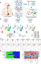
          The tumor microenvironment is a highly complex ecosystem of diverse cell types, which shape cancer biology and impact the responsiveness to therapy. Here, we analyze the microenvironment of esophageal squamous cell carcinoma (ESCC) using single-cell transcriptome sequencing in 62,161 cells from blood, adjacent nonmalignant and matched tumor samples from 11 ESCC patients. We uncover heterogeneity in most cell types of the ESCC stroma, particularly in the fibroblast and immune cell compartments. We identify a tumor-specific subset of CST1 + myofibroblasts with prognostic values and potential biological significance. CST1 + myofibroblasts are also highly tumor-specific in other cancer types. Additionally, a subset of antigen-presenting fibroblasts is revealed and validated. Analyses of myeloid and T lymphoid lineages highlight the immunosuppressive nature of the ESCC microenvironment, and identify cancer-specific expression of immune checkpoint inhibitors. This work establishes a rich resource of stromal cell types of the ESCC microenvironment for further understanding of ESCC biology.

          Abstract

          The microenvironment of oesophageal squamous cell carcinomas (ESCC) is heterogeneous and can strongly impact response to treatment. Here, the authors characterize the ESCC tumour microenvironment with single-cell RNA-seq, finding CST1 + myofibroblasts with potential biological and prognostic significance as well as immunosuppression signatures.

          Related collections

          Most cited references65

          • Record: found
          • Abstract: found
          • Article: not found

          Gene set enrichment analysis: A knowledge-based approach for interpreting genome-wide expression profiles

          Although genomewide RNA expression analysis has become a routine tool in biomedical research, extracting biological insight from such information remains a major challenge. Here, we describe a powerful analytical method called Gene Set Enrichment Analysis (GSEA) for interpreting gene expression data. The method derives its power by focusing on gene sets, that is, groups of genes that share common biological function, chromosomal location, or regulation. We demonstrate how GSEA yields insights into several cancer-related data sets, including leukemia and lung cancer. Notably, where single-gene analysis finds little similarity between two independent studies of patient survival in lung cancer, GSEA reveals many biological pathways in common. The GSEA method is embodied in a freely available software package, together with an initial database of 1,325 biologically defined gene sets.
            Bookmark
            • Record: found
            • Abstract: found
            • Article: not found

            Comprehensive Integration of Single-Cell Data

            Single-cell transcriptomics has transformed our ability to characterize cell states, but deep biological understanding requires more than a taxonomic listing of clusters. As new methods arise to measure distinct cellular modalities, a key analytical challenge is to integrate these datasets to better understand cellular identity and function. Here, we develop a strategy to "anchor" diverse datasets together, enabling us to integrate single-cell measurements not only across scRNA-seq technologies, but also across different modalities. After demonstrating improvement over existing methods for integrating scRNA-seq data, we anchor scRNA-seq experiments with scATAC-seq to explore chromatin differences in closely related interneuron subsets and project protein expression measurements onto a bone marrow atlas to characterize lymphocyte populations. Lastly, we harmonize in situ gene expression and scRNA-seq datasets, allowing transcriptome-wide imputation of spatial gene expression patterns. Our work presents a strategy for the assembly of harmonized references and transfer of information across datasets.
              Bookmark
              • Record: found
              • Abstract: found
              • Article: not found

              SCENIC: Single-cell regulatory network inference and clustering

              Although single-cell RNA-seq is revolutionizing biology, data interpretation remains a challenge. We present SCENIC for the simultaneous reconstruction of gene regulatory networks and identification of cell states. We apply SCENIC to a compendium of single-cell data from tumors and brain, and demonstrate that the genomic regulatory code can be exploited to guide the identification of transcription factors and cell states. SCENIC provides critical biological insights into the mechanisms driving cellular heterogeneity.
                Bookmark

                Author and article information

                Contributors
                huy.dinh@wisc.edu
                lyxu@stu.edu.cn
                dchlin11@gmail.com
                nmli@stu.edu.cn
                Journal
                Nat Commun
                Nat Commun
                Nature Communications
                Nature Publishing Group UK (London )
                2041-1723
                17 December 2021
                17 December 2021
                2021
                : 12
                : 7335
                Affiliations
                [1 ]GRID grid.14003.36, ISNI 0000 0001 2167 3675, McArdle Laboratory for Cancer Research, , University of Wisconsin-Madison School of Medicine and Public Health, ; Madison, WI USA
                [2 ]GRID grid.185006.a, ISNI 0000 0004 0461 3162, Division of Inflammation Biology, , La Jolla Institute for Immunology, ; La Jolla, CA USA
                [3 ]GRID grid.411679.c, ISNI 0000 0004 0605 3373, Guangdong Provincial Key Laboratory of Infectious Diseases and Molecular Immunopathology, The Key Laboratory of Molecular Biology for High Cancer Incidence Coastal Chaoshan Area, , Shantou University Medical College, ; Shantou, China
                [4 ]Guangdong Esophageal Cancer Research Institute, Shantou Sub-center, Shantou, China
                [5 ]GRID grid.411917.b, Department of Thoracic Surgery, , Cancer Hospital of Shantou University Medical College, ; Shantou, China
                [6 ]GRID grid.452734.3, Shantou Central Hospital, ; Shantou, China
                [7 ]GRID grid.50956.3f, ISNI 0000 0001 2152 9905, Department of Medicine, , Samuel Oschin Comprehensive Cancer Institute, Cedars-Sinai Medical Center, ; Los Angeles, CA USA
                [8 ]GRID grid.19006.3e, ISNI 0000 0000 9632 6718, Department of Obstetrics and Gynecology and Samuel Oschin Comprehensive Cancer Institute, , Cedars-Sinai Medical Center, David Geffen School of Medicine at UCLA, ; Los Angeles, CA USA
                [9 ]GRID grid.50956.3f, ISNI 0000 0001 2152 9905, Board of Governors Regenerative Medicine Institute and Department of Biomedical Sciences, , Cedars-Sinai Medical Center, ; Los Angeles, CA USA
                Author information
                http://orcid.org/0000-0002-3307-1126
                http://orcid.org/0000-0003-1594-8884
                http://orcid.org/0000-0001-5119-8721
                http://orcid.org/0000-0002-3735-3390
                http://orcid.org/0000-0001-5839-9913
                http://orcid.org/0000-0002-1618-4292
                http://orcid.org/0000-0002-1951-367X
                http://orcid.org/0000-0001-6375-3614
                Article
                27599
                10.1038/s41467-021-27599-5
                8683407
                34921160
                2cfe965f-28ba-4264-8f51-05ee2060586a
                © The Author(s) 2021

                Open Access This article is licensed under a Creative Commons Attribution 4.0 International License, which permits use, sharing, adaptation, distribution and reproduction in any medium or format, as long as you give appropriate credit to the original author(s) and the source, provide a link to the Creative Commons license, and indicate if changes were made. The images or other third party material in this article are included in the article’s Creative Commons license, unless indicated otherwise in a credit line to the material. If material is not included in the article’s Creative Commons license and your intended use is not permitted by statutory regulation or exceeds the permitted use, you will need to obtain permission directly from the copyright holder. To view a copy of this license, visit http://creativecommons.org/licenses/by/4.0/.

                History
                : 9 June 2020
                : 29 November 2021
                Funding
                Funded by: FundRef https://doi.org/10.13039/100000002, U.S. Department of Health & Human Services | National Institutes of Health (NIH);
                Award ID: R01 CA202987
                Award ID: U01 CA224766
                Award Recipient :
                Funded by: U.S. Department of Health & Human Services | National Institutes of Health (NIH)
                Funded by: FundRef https://doi.org/10.13039/501100003453, Natural Science Foundation of Guangdong Province (Guangdong Natural Science Foundation);
                Award ID: No. U1601229
                Award Recipient :
                Categories
                Article
                Custom metadata
                © The Author(s) 2021

                Uncategorized
                cancer microenvironment,oesophageal cancer,cancer genomics,tumour heterogeneity,transcriptomics

                Comments

                Comment on this article