185
views
0
recommends
+1 Recommend
0 collections
    16
    shares
      • Record: found
      • Abstract: found
      • Article: found
      Is Open Access

      Implications of the Hybrid Epithelial/Mesenchymal Phenotype in Metastasis

      review-article

      Read this article at

      Bookmark
          There is no author summary for this article yet. Authors can add summaries to their articles on ScienceOpen to make them more accessible to a non-specialist audience.

          Abstract

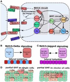
          Transitions between epithelial and mesenchymal phenotypes – the epithelial to ­mesenchymal transition (EMT) and its reverse the mesenchymal to epithelial transition (MET) – are hallmarks of cancer metastasis. While transitioning between the epithelial and mesenchymal phenotypes, cells can also attain a hybrid epithelial/mesenchymal (E/M) (i.e., partial or intermediate EMT) phenotype. Cells in this phenotype have mixed epithelial (e.g., adhesion) and mesenchymal (e.g., migration) properties, thereby allowing them to move collectively as clusters. If these clusters reach the bloodstream intact, they can give rise to clusters of circulating tumor cells (CTCs), as have often been seen experimentally. Here, we review the operating principles of the core regulatory network for EMT/MET that acts as a “three-way” switch giving rise to three distinct phenotypes – E, M and hybrid E/M – and present a theoretical framework that can elucidate the role of many other players in regulating epithelial plasticity. Furthermore, we highlight recent studies on partial EMT and its association with drug resistance and tumor-initiating potential; and discuss how cell–cell communication between cells in a partial EMT phenotype can enable the formation of clusters of CTCs. These clusters can be more apoptosis-resistant and have more tumor-initiating potential than singly moving CTCs with a wholly mesenchymal (complete EMT) phenotype. Also, more such clusters can be formed under inflammatory conditions that are often generated by various therapies. Finally, we discuss the multiple advantages that the partial EMT or hybrid E/M phenotype have as compared to a complete EMT phenotype and argue that these collectively migrating cells are the primary “bad actors” of metastasis.

          Related collections

          Most cited references144

          • Record: found
          • Abstract: found
          • Article: not found

          Identification of pancreatic cancer stem cells.

          Emerging evidence has suggested that the capability of a tumor to grow and propagate is dependent on a small subset of cells within a tumor, termed cancer stem cells. Although data have been provided to support this theory in human blood, brain, and breast cancers, the identity of pancreatic cancer stem cells has not been determined. Using a xenograft model in which primary human pancreatic adenocarcinomas were grown in immunocompromised mice, we identified a highly tumorigenic subpopulation of pancreatic cancer cells expressing the cell surface markers CD44, CD24, and epithelial-specific antigen (ESA). Pancreatic cancer cells with the CD44(+)CD24(+)ESA(+) phenotype (0.2-0.8% of pancreatic cancer cells) had a 100-fold increased tumorigenic potential compared with nontumorigenic cancer cells, with 50% of animals injected with as few as 100 CD44(+)CD24(+)ESA(+) cells forming tumors that were histologically indistinguishable from the human tumors from which they originated. The enhanced ability of CD44(+)CD24(+)ESA(+) pancreatic cancer cells to form tumors was confirmed in an orthotopic pancreatic tail injection model. The CD44(+)CD24(+)ESA(+) pancreatic cancer cells showed the stem cell properties of self-renewal, the ability to produce differentiated progeny, and increased expression of the developmental signaling molecule sonic hedgehog. Identification of pancreatic cancer stem cells and further elucidation of the signaling pathways that regulate their growth and survival may provide novel therapeutic approaches to treat pancreatic cancer, which is notoriously resistant to standard chemotherapy and radiation.
            Bookmark
            • Record: found
            • Abstract: found
            • Article: found
            Is Open Access

            Plasticity of cell migration: a multiscale tuning model

            Cell migration underlies tissue formation, maintenance, and regeneration as well as pathological conditions such as cancer invasion. Structural and molecular determinants of both tissue environment and cell behavior define whether cells migrate individually (through amoeboid or mesenchymal modes) or collectively. Using a multiparameter tuning model, we describe how dimension, density, stiffness, and orientation of the extracellular matrix together with cell determinants—including cell–cell and cell–matrix adhesion, cytoskeletal polarity and stiffness, and pericellular proteolysis—interdependently control migration mode and efficiency. Motile cells integrate variable inputs to adjust interactions among themselves and with the matrix to dictate the migration mode. The tuning model provides a matrix of parameters that control cell movement as an adaptive and interconvertible process with relevance to different physiological and pathological contexts.
              Bookmark
              • Record: found
              • Abstract: found
              • Article: found
              Is Open Access

              Human breast cancer cell lines contain stem-like cells that self-renew, give rise to phenotypically diverse progeny and survive chemotherapy

              Introduction The phenotypic and functional differences between cells that initiate human breast tumors (cancer stem cells) and those that comprise the tumor bulk are difficult to study using only primary tumor tissue. We embarked on this study hypothesizing that breast cancer cell lines would contain analogous hierarchical differentiation programs to those found in primary breast tumors. Methods Eight human breast cell lines (human mammary epithelial cells, and MCF10A, MCF7, SUM149, SUM159, SUM1315 and MDA.MB.231 cells) were analyzed using flow cytometry for CD44, CD24, and epithelial-specific antigen (ESA) expression. Limiting dilution orthotopic injections were used to evaluate tumor initiation, while serial colony-forming unit, reconstitution and tumorsphere assays were performed to assess self-renewal and differentiation. Pulse-chase bromodeoxyuridine (5-bromo-2-deoxyuridine [BrdU]) labeling was used to examine cell cycle and label-retention of cancer stem cells. Cells were treated with paclitaxol and 5-fluorouracil to test selective resistance to chemotherapy, and gene expression profile after chemotherapy were examined. Results The percentage of CD44+/CD24- cells within cell lines does not correlate with tumorigenicity, but as few as 100 cells can form tumors when sorted for CD44+/CD24-/low/ESA+. Furthermore, CD44+/CD24-/ESA+ cells can self-renew, reconstitute the parental cell line, retain BrdU label, and preferentially survive chemotherapy. Conclusion These data validate the use of cancer cell lines as models for the development and testing of novel therapeutics aimed at eradicating cancer stem cells.
                Bookmark

                Author and article information

                Contributors
                Journal
                Front Oncol
                Front Oncol
                Front. Oncol.
                Frontiers in Oncology
                Frontiers Media S.A.
                2234-943X
                20 July 2015
                2015
                : 5
                : 155
                Affiliations
                [1] 1Center for Theoretical Biological Physics, Rice University , Houston, TX, USA
                [2] 2Department of Bioengineering, Rice University , Houston, TX, USA
                [3] 3Institute of Physics, University of São Paulo , São Paulo, Brazil
                [4] 4Department of Chemistry, Rice University , Houston, TX, USA
                [5] 5Graduate Program in Systems, Synthetic and Physical Biology, Rice University , Houston, TX, USA
                [6] 6School of Physics and Astronomy, and The Sagol School of Neuroscience, Tel-Aviv University , Tel-Aviv, Israel
                [8] 7Department of Biosciences, Rice University , Houston, TX, USA
                [7] 8Department of Physics and Astronomy, Rice University , Houston, TX, USA
                Author notes

                Edited by: Lars Bolund, Aarhus University, Denmark

                Reviewed by: Shree Ram Singh, National Cancer Institute, USA; Hiroyuki Moriyama, Kinki University, Japan

                *Correspondence: José N. Onuchic and Herbert Levine, Bioscience Research Collaborative, Rice University, 6100 Main Street, MS 654, Houston, TX 77005-1827, USA, jonuchic@ 123456rice.edu ; herbert.levine@ 123456rice.edu

                Deceased on June 5, 2015.

                Specialty section: This article was submitted to Stem Cell Research, a section of the journal Frontiers in Oncology

                Article
                10.3389/fonc.2015.00155
                4507461
                26258068
                2cd3e675-1ff1-4e7e-8cf0-5a69aa11f6c5
                Copyright © 2015 Jolly, Boareto, Huang, Jia, Lu, Ben-Jacob, Onuchic and Levine.

                This is an open-access article distributed under the terms of the Creative Commons Attribution License (CC BY). The use, distribution or reproduction in other forums is permitted, provided the original author(s) or licensor are credited and that the original publication in this journal is cited, in accordance with accepted academic practice. No use, distribution or reproduction is permitted which does not comply with these terms.

                History
                : 22 May 2015
                : 29 June 2015
                Page count
                Figures: 9, Tables: 0, Equations: 0, References: 230, Pages: 19, Words: 16633
                Funding
                Funded by: National Science Foundation (NSF) Center for Theoretical Biological Physics
                Award ID: NSF PHY-1427654
                Funded by: NSF
                Award ID: MCB-1214457
                Funded by: CPRIT (Cancer Prevention and Research Institute of Texas) Scholar in Cancer Research of the State of Texas at Rice University
                Funded by: CPRIT
                Funded by: Tauber Family Funds and the Maguy-Glass Chair in Physics of Complex Systems
                Funded by: FAPESP
                Award ID: 2013/14438-8
                Funded by: Keck Center for Interdisciplinary Bioscience Training of the Gulf Coast Consortia
                Funded by: CPRIT
                Award ID: RP140113
                Categories
                Oncology
                Review

                Oncology & Radiotherapy
                partial emt,intermediate emt,cancer stem cells,cell-fate decisions,cancer systems biology

                Comments

                Comment on this article