38
views
0
recommends
+1 Recommend
0 collections
    0
    shares
      • Record: found
      • Abstract: found
      • Article: found
      Is Open Access

      Musashi1 regulates breast tumor cell proliferation and is a prognostic indicator of poor survival

      research-article

      Read this article at

      Bookmark
          There is no author summary for this article yet. Authors can add summaries to their articles on ScienceOpen to make them more accessible to a non-specialist audience.

          Abstract

          Background

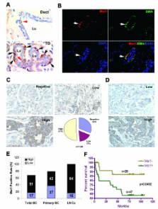
          Musashi1 (Msi1) is a conserved RNA-binding protein that regulates the Notch and Wnt pathways, and serves as a stem cell marker in the breast and other tissues. It is unknown how Msi1 relates to other breast cancer markers, whether it denotes tumor initiating cells (TICs), and how it affects gene expression and tumor cell survival in breast cancer cells.

          Results

          Msi1 expression was analyzed in 20 breast cancer cell lines and in 140 primary breast tumors by western blotting and immunohistochemistry, respectively. Lentivirus RNA interference was used to reduce Msi1 expression in breast cancer cell lines MCF-7 and T47D grown as spheroid cultures and to assess stem cell gene expression and the growth of these cell lines as xenografts. In normal human breast tissue, Msi1 was expressed in 10.6% of myoepithelum and 1.2% of ductal epithelium in the terminal ductal lobular unit (TDLU), whereas, less than 0.05% of ductal epithelium and myoepithelium in large ducts outside the TDLU expressed Msi1. Msi1 was expressed in 55% of the breast cancer cell lines and correlated with ErbB2 expression in 50% of the cell lines. Msi1 was expressed in 68% of primary tumors and in 100% of lymph node metastases, and correlated with 5 year survival. Msi1 was enriched in CD133 + MCF-7 and T47D cells and in spheroid cultures of these cells, and Msi1 'knockdown' (KD) with a lentivirus-expressed shRNA decreased the number and size of spheroid colonies. Msi1 KD reduced Notch1, c-Myc, ErbB2 and pERK1/2 expression, and increased p21 CIP1 expression, which is consistent with known Msi1 target mRNAs. Msi1 KD also reduced the expression of the somatic and embryonic stem cell markers, CD133, Bmi1, Sox2, Nanog and Oct4. Xenografts of MCF-7 and T47D Msi1 KD cells resulted in a marked reduction of tumor growth, reduced Msi1 and Notch1 expression and increased p21 CIP1 expression.

          Conclusion

          Msi1 is a negative prognostic indicator of breast cancer patient survival, and is indicative of tumor cells with stem cell-like characteristics. Msi1 KD reduces tumor cell survival and tumor xenograft growth, suggesting that it may represent a novel target for drug discovery.

          Related collections

          Most cited references30

          • Record: found
          • Abstract: found
          • Article: not found

          Cancer Statistics, 2008

          Each year, the American Cancer Society estimates the number of new cancer cases and deaths expected in the United States in the current year and compiles the most recent data on cancer incidence, mortality, and survival based on incidence data from the National Cancer Institute, Centers for Disease Control and Prevention, and the North American Association of Central Cancer Registries and mortality data from the National Center for Health Statistics. Incidence and death rates are age-standardized to the 2000 US standard million population. A total of 1,437,180 new cancer cases and 565,650 deaths from cancer are projected to occur in the United States in 2008. Notable trends in cancer incidence and mortality include stabilization of incidence rates for all cancer sites combined in men from 1995 through 2004 and in women from 1999 through 2004 and a continued decrease in the cancer death rate since 1990 in men and since 1991 in women. Overall cancer death rates in 2004 compared with 1990 in men and 1991 in women decreased by 18.4% and 10.5%, respectively, resulting in the avoidance of over a half million deaths from cancer during this time interval. This report also examines cancer incidence, mortality, and survival by site, sex, race/ethnicity, education, geographic area, and calendar year, as well as the proportionate contribution of selected sites to the overall trends. Although much progress has been made in reducing mortality rates, stabilizing incidence rates, and improving survival, cancer still accounts for more deaths than heart disease in persons under age 85 years. Further progress can be accelerated by supporting new discoveries and by applying existing cancer control knowledge across all segments of the population.
            Bookmark
            • Record: found
            • Abstract: found
            • Article: not found

            The response of CD24(-/low)/CD44+ breast cancer-initiating cells to radiation.

            If cancer arises and is maintained by a small population of cancer-initiating cells within every tumor, understanding how these cells react to cancer treatment will facilitate improvement of cancer treatment in the future. Cancer-initiating cells can now be prospectively isolated from breast cancer cell lines and tumor samples and propagated as mammospheres in vitro under serum-free conditions. CD24(-/low)/CD44+ cancer-initiating cells were isolated from MCF-7 and MDA-MB-231 breast cancer monolayer cultures and propagated as mammospheres. Their response to radiation was investigated by assaying clonogenic survival and by measuring reactive oxygen species (ROS) levels, phosphorylation of the replacement histone H2AX, CD44 levels, CD24 levels, and Notch-1 activation using flow cytometry. All statistical tests were two-sided. Cancer-initiating cells were more resistant to radiation than cells grown as monolayer cultures (MCF-7: monolayer cultures, mean surviving fraction at 2 Gy [SF(2Gy)] = 0.2, versus mammospheres, mean SF(2Gy) = 0.46, difference = 0.26, 95% confidence interval [CI] = 0.05 to 0.47; P = .026; MDA-MB-231: monolayer cultures, mean SF(2Gy) = 0.5, versus mammospheres, mean SF(2Gy) = 0.69, difference = 0.19, 95% CI = -0.07 to 0.45; P = .09). Levels of ROS increased in both mammospheres and monolayer cultures after irradiation with a single dose of 10 Gy but were lower in mammospheres than in monolayer cultures (MCF-7 monolayer cultures: 0 Gy, mean = 1.0, versus 10 Gy, mean = 3.32, difference = 2.32, 95% CI = 0.67 to 3.98; P = .026; mammospheres: 0 Gy, mean = 0.58, versus 10 Gy, mean = 1.46, difference = 0.88, 95% CI = 0.20 to 1.56; P = .031); phosphorylation of H2AX increased in irradiated monolayer cultures, but no change was observed in mammospheres. Fractionated doses of irradiation increased activation of Notch-1 (untreated, mean = 10.7, versus treated, mean = 15.1, difference = 4.4, 95% CI = 2.7 to 6.1, P = .002) and the percentage of the cancer stem/initiating cells in the nonadherent cell population of MCF-7 monolayer cultures (untreated, mean = 3.52%, versus treated, mean = 7.5%, difference = 3.98%, 95% CI = 1.67% to 6.25%, P = .009). Breast cancer-initiating cells are a relatively radioresistant subpopulation of breast cancer cells and increase in numbers after short courses of fractionated irradiation. These findings offer a possible mechanism for the accelerated repopulation of tumor cells observed during gaps in radiotherapy.
              Bookmark
              • Record: found
              • Abstract: found
              • Article: not found

              CD133(+) and CD133(-) glioblastoma-derived cancer stem cells show differential growth characteristics and molecular profiles.

              Although glioblastomas show the same histologic phenotype, biological hallmarks such as growth and differentiation properties vary considerably between individual cases. To investigate whether different subtypes of glioblastomas might originate from different cells of origin, we cultured tumor cells from 22 glioblastomas under medium conditions favoring the growth of neural and cancer stem cells (CSC). Secondary glioblastoma (n = 7)-derived cells did not show any growth in the medium used, suggesting the absence of neural stem cell-like tumor cells. In contrast, 11/15 primary glioblastomas contained a significant CD133(+) subpopulation that displayed neurosphere-like, nonadherent growth and asymmetrical cell divisions yielding cells expressing markers characteristic for all three neural lineages. Four of 15 cell lines derived from primary glioblastomas grew adherently in vitro and were driven by CD133(-) tumor cells that fulfilled stem cell criteria. Both subtypes were similarly tumorigenic in nude mice in vivo. Clinically, CD133(-) glioblastomas were characterized by a lower proliferation index, whereas glial fibrillary acidic protein staining was similar. GeneArray analysis revealed 117 genes to be differentially expressed by these two subtypes. Together, our data provide first evidence that CD133(+) CSC maintain only a subset of primary glioblastomas. The remainder stems from previously unknown CD133(-) tumor cells with apparent stem cell-like properties but distinct molecular profiles and growth characteristics in vitro and in vivo.
                Bookmark

                Author and article information

                Journal
                Mol Cancer
                Molecular Cancer
                BioMed Central
                1476-4598
                2010
                21 August 2010
                : 9
                : 221
                Affiliations
                [1 ]Department of Oncology, Georgetown University, and Lombardi Comprehensive Cancer Center, Washington, DC 20007, USA
                [2 ]Cell and Cancer Biology Branch, Center for Cancer Research, National Cancer Institute, National Institutes of Health, Bethesda, MD 20892, USA
                [3 ]Department of Cell and Structural Biology, University of Texas Health Science Center at San Antonio, TX, USA
                [4 ]Department of Epidemiology, State Key Lab of Respiratory Disease, Guangzhou Medical College, Guangzhou, Guangdong, China
                [5 ]Department of Physiology, Keio University School of Medicine, Tokyo, Japan
                Article
                1476-4598-9-221
                10.1186/1476-4598-9-221
                2939568
                20727204
                2c97e7c0-bd5f-4349-9721-29112f282fc4
                Copyright ©2010 Wang et al; licensee BioMed Central Ltd.

                This is an Open Access article distributed under the terms of the Creative Commons Attribution License ( http://creativecommons.org/licenses/by/2.0), which permits unrestricted use, distribution, and reproduction in any medium, provided the original work is properly cited.

                History
                : 5 April 2010
                : 21 August 2010
                Categories
                Research

                Oncology & Radiotherapy
                Oncology & Radiotherapy

                Comments

                Comment on this article