14
views
0
recommends
+1 Recommend
0 collections
    0
    shares
      • Record: found
      • Abstract: found
      • Article: not found

      Mitochondrial complex III is necessary for endothelial cell proliferation during angiogenesis

      research-article

      Read this article at

      ScienceOpenPublisherPMC
      Bookmark
          There is no author summary for this article yet. Authors can add summaries to their articles on ScienceOpen to make them more accessible to a non-specialist audience.

          Abstract

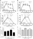
          Endothelial cells (ECs) require glycolysis for proliferation and migration during angiogenesis; however, the necessity for the mitochondrial respiratory chain during angiogenesis is not known. Here we report that inhibition of respiratory chain complex III impairs proliferation, but not migration of ECs in vitro by decreasing the NAD+/NADH ratio. To determine whether mitochondrial respiration is necessary for angiogenesis in vivo, we conditionally ablate a subunit of the respiratory chain complex III (QPC) in ECs. Loss of QPC decreases respiration, resulting in diminished EC proliferation, and impairment in retinal and tumor angiogenesis. Loss of QPC does not decrease genes associated with anabolism or nucleotides levels in ECs, but diminishes amino acid levels. Our findings indicate that mitochondrial respiration is necessary for angiogenesis, and that the primary role of mitochondria in ECs is to serve as biosynthetic organelles for cell proliferation.

          Related collections

          Most cited references36

          • Record: found
          • Abstract: found
          • Article: found
          Is Open Access

          Metformin inhibits mitochondrial complex I of cancer cells to reduce tumorigenesis

          Recent epidemiological and laboratory-based studies suggest that the anti-diabetic drug metformin prevents cancer progression. How metformin diminishes tumor growth is not fully understood. In this study, we report that in human cancer cells, metformin inhibits mitochondrial complex I (NADH dehydrogenase) activity and cellular respiration. Metformin inhibited cellular proliferation in the presence of glucose, but induced cell death upon glucose deprivation, indicating that cancer cells rely exclusively on glycolysis for survival in the presence of metformin. Metformin also reduced hypoxic activation of hypoxia-inducible factor 1 (HIF-1). All of these effects of metformin were reversed when the metformin-resistant Saccharomyces cerevisiae NADH dehydrogenase NDI1 was overexpressed. In vivo, the administration of metformin to mice inhibited the growth of control human cancer cells but not those expressing NDI1. Thus, we have demonstrated that metformin's inhibitory effects on cancer progression are cancer cell autonomous and depend on its ability to inhibit mitochondrial complex I. DOI: http://dx.doi.org/10.7554/eLife.02242.001
            Bookmark
            • Record: found
            • Abstract: found
            • Article: not found

            A general introduction to the biochemistry of mitochondrial fatty acid β-oxidation

            Over the years, the mitochondrial fatty acid β-oxidation (FAO) pathway has been characterised at the biochemical level as well as the molecular biological level. FAO plays a pivotal role in energy homoeostasis, but it competes with glucose as the primary oxidative substrate. The mechanisms behind this so-called glucose–fatty acid cycle operate at the hormonal, transcriptional and biochemical levels. Inherited defects for most of the FAO enzymes have been identified and characterised and are currently included in neonatal screening programmes. Symptoms range from hypoketotic hypoglycaemia to skeletal and cardiac myopathies. The pathophysiology of these diseases is still not completely understood, hampering optimal treatment. Studies of patients and mouse models will contribute to our understanding of the pathogenesis and will ultimately lead to better treatment.
              Bookmark
              • Record: found
              • Abstract: found
              • Article: not found

              Inhibition of the Glycolytic Activator PFKFB3 in Endothelium Induces Tumor Vessel Normalization, Impairs Metastasis, and Improves Chemotherapy.

              Abnormal tumor vessels promote metastasis and impair chemotherapy. Hence, tumor vessel normalization (TVN) is emerging as an anti-cancer treatment. Here, we show that tumor endothelial cells (ECs) have a hyper-glycolytic metabolism, shunting intermediates to nucleotide synthesis. EC haplo-deficiency or blockade of the glycolytic activator PFKFB3 did not affect tumor growth, but reduced cancer cell invasion, intravasation, and metastasis by normalizing tumor vessels, which improved vessel maturation and perfusion. Mechanistically, PFKFB3 inhibition tightened the vascular barrier by reducing VE-cadherin endocytosis in ECs, and rendering pericytes more quiescent and adhesive (via upregulation of N-cadherin) through glycolysis reduction; it also lowered the expression of cancer cell adhesion molecules in ECs by decreasing NF-κB signaling. PFKFB3-blockade treatment also improved chemotherapy of primary and metastatic tumors.
                Bookmark

                Author and article information

                Journal
                101736592
                48119
                Nat Metab
                Nat Metab
                Nature metabolism
                2522-5812
                5 November 2018
                7 January 2019
                January 2019
                07 July 2019
                : 1
                : 1
                : 158-171
                Affiliations
                [1 ]Departments of Medicine and Robert H. Lurie Cancer Center, Northwestern University Feinberg School of Medicine, Chicago, IL 60611
                [2 ]Center for Vascular and Developmental Biology, Feinberg Cardiovascular Research Institute, Northwestern University, Chicago, IL 60611
                [3 ]Robert H. Lurie Cancer Center Metabolomics Core, Northwestern University Feinberg School of Medicine, Chicago, IL 60611
                [4 ]Department of Biochemistry and Molecular Genetics, Northwestern University Feinberg School of Medicine, Chicago, IL 60611
                Author notes

                AUTHOR CONTRIBUIONS

                L.D. carried out the majority of experiments and data analysis. P.G. performed the HPLC-MS/MS metabolite analysis. C.A.M. analyzed the RNA sequencing data. H.J.G. and S.E.W. provided intellectual and technical expertise. L.D. and N.S.C. provided intellectual input and wrote the paper.

                [* ]Corresponding author: nav@ 123456northwestern.edu
                Article
                NIHMS1511426
                10.1038/s42255-018-0011-x
                6521885
                31106291
                2af55e72-2046-426b-b599-a143918980df

                Users may view, print, copy, and download text and data-mine the content in such documents, for the purposes of academic research, subject always to the full Conditions of use: http://www.nature.com/authors/editorial_policies/license.html#terms

                History
                Categories
                Article

                Comments

                Comment on this article