56
views
0
recommends
+1 Recommend
0 collections
    0
    shares
      • Record: found
      • Abstract: found
      • Article: not found

      Melanopsin-Driven Light Adaptation in Mouse Vision

      research-article

      Read this article at

      ScienceOpenPublisherPMC
      Bookmark
          There is no author summary for this article yet. Authors can add summaries to their articles on ScienceOpen to make them more accessible to a non-specialist audience.

          Summary

          Background

          In bright light, mammals use a distinct photopigment (melanopsin) to measure irradiance for centrally mediated responses such as circadian entrainment. We aimed to determine whether the information generated by melanopsin is also used by the visual system as a signal for light adaptation. To this end, we compared retinal and thalamic responses to a range of artificial and natural visual stimuli presented using spectral compositions that either approximate the mouse’s experience of natural daylight (“daylight”) or are selectively depleted of wavelengths to which melanopsin is most sensitive (“mel-low”).

          Results

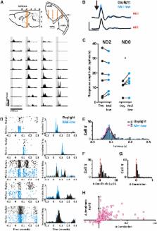
          We found reproducible and reversible changes in the flash electroretinogram between daylight and mel-low. Simultaneous recording in the dorsal lateral geniculate nucleus (dLGN) revealed that these reflect changes in feature selectivity of visual circuits in both temporal and spatial dimensions. A substantial fraction of units preferred finer spatial patterns in the daylight condition, while the population of direction-sensitive units became tuned to faster motion. The dLGN contained a richer, more reliable encoding of natural scenes in the daylight condition. These effects were absent in mice lacking melanopsin.

          Conclusions

          The feature selectivity of many neurons in the mouse dLGN is adjusted according to a melanopsin-dependent measure of environmental brightness. These changes originate, at least in part, within the retina. Melanopsin performs a role analogous to a photographer’s light meter, providing an independent measure of irradiance that determines optimal setting for visual circuits.

          Highlights

          • Adjustments in visual performance in daylight conditions are driven by melanopsin

          • Melanopsin adjusts visual feature selectivity in spatial and temporal dimensions

          • Melanopsin facilitates a richer and more reliable encoding of natural scenes

          Abstract

          Allen et al. show that aspects of light adaptation rely on a measure of brightness provided by melanopsin, the retina’s most recently discovered photoreceptor. Recordings from the mouse thalamus show that melanopsin controls the types of visual features that excite single neurons, allowing a richer, more reliable representation of natural scenes.

          Related collections

          Most cited references43

          • Record: found
          • Abstract: found
          • Article: not found

          Melanopsin-expressing ganglion cells in primate retina signal colour and irradiance and project to the LGN.

          Human vision starts with the activation of rod photoreceptors in dim light and short (S)-, medium (M)-, and long (L)- wavelength-sensitive cone photoreceptors in daylight. Recently a parallel, non-rod, non-cone photoreceptive pathway, arising from a population of retinal ganglion cells, was discovered in nocturnal rodents. These ganglion cells express the putative photopigment melanopsin and by signalling gross changes in light intensity serve the subconscious, 'non-image-forming' functions of circadian photoentrainment and pupil constriction. Here we show an anatomically distinct population of 'giant', melanopsin-expressing ganglion cells in the primate retina that, in addition to being intrinsically photosensitive, are strongly activated by rods and cones, and display a rare, S-Off, (L + M)-On type of colour-opponent receptive field. The intrinsic, rod and (L + M) cone-derived light responses combine in these giant cells to signal irradiance over the full dynamic range of human vision. In accordance with cone-based colour opponency, the giant cells project to the lateral geniculate nucleus, the thalamic relay to primary visual cortex. Thus, in the diurnal trichromatic primate, 'non-image-forming' and conventional 'image-forming' retinal pathways are merged, and the melanopsin-based signal might contribute to conscious visual perception.
            Bookmark
            • Record: found
            • Abstract: found
            • Article: not found

            Melanopsin and rod-cone photoreceptive systems account for all major accessory visual functions in mice.

            In the mammalian retina, besides the conventional rod-cone system, a melanopsin-associated photoreceptive system exists that conveys photic information for accessory visual functions such as pupillary light reflex and circadian photo-entrainment. On ablation of the melanopsin gene, retinal ganglion cells that normally express melanopsin are no longer intrinsically photosensitive. Furthermore, pupil reflex, light-induced phase delays of the circadian clock and period lengthening of the circadian rhythm in constant light are all partially impaired. Here, we investigated whether additional photoreceptive systems participate in these responses. Using mice lacking rods and cones, we measured the action spectrum for phase-shifting the circadian rhythm of locomotor behaviour. This spectrum matches that for the pupillary light reflex in mice of the same genotype, and that for the intrinsic photosensitivity of the melanopsin-expressing retinal ganglion cells. We have also generated mice lacking melanopsin coupled with disabled rod and cone phototransduction mechanisms. These animals have an intact retina but fail to show any significant pupil reflex, to entrain to light/dark cycles, and to show any masking response to light. Thus, the rod-cone and melanopsin systems together seem to provide all of the photic input for these accessory visual functions.
              Bookmark
              • Record: found
              • Abstract: found
              • Article: not found

              Melanopsin is required for non-image-forming photic responses in blind mice.

              Although mice lacking rod and cone photoreceptors are blind, they retain many eye-mediated responses to light, possibly through photosensitive retinal ganglion cells. These cells express melanopsin, a photopigment that confers this photosensitivity. Mice lacking melanopsin still retain nonvisual photoreception, suggesting that rods and cones could operate in this capacity. We observed that mice with both outer-retinal degeneration and a deficiency in melanopsin exhibited complete loss of photoentrainment of the circadian oscillator, pupillary light responses, photic suppression of arylalkylamine-N-acetyltransferase transcript, and acute suppression of locomotor activity by light. This indicates the importance of both nonvisual and classical visual photoreceptor systems for nonvisual photic responses in mammals.
                Bookmark

                Author and article information

                Contributors
                Journal
                Curr Biol
                Curr. Biol
                Current Biology
                Cell Press
                0960-9822
                1879-0445
                03 November 2014
                03 November 2014
                : 24
                : 21
                : 2481-2490
                Affiliations
                [1 ]Faculty of Life Sciences, Manchester University, Oxford Road, Manchester M13 9PT, UK
                Author notes
                []Corresponding author annette.allen@ 123456manchester.ac.uk
                [∗∗ ]Corresponding author robert.lucas@ 123456manchester.ac.uk
                Article
                S0960-9822(14)01135-X
                10.1016/j.cub.2014.09.015
                4228053
                25308073
                299d516f-539d-492b-b76c-2ab676c59e8d
                © 2014 The Authors
                History
                : 12 June 2014
                : 6 August 2014
                : 3 September 2014
                Categories
                Article

                Life sciences
                Life sciences

                Comments

                Comment on this article