6
views
0
recommends
+1 Recommend
0 collections
    0
    shares
      • Record: found
      • Abstract: found
      • Article: found
      Is Open Access

      Methylglyoxal, a glycolysis metabolite, triggers metastasis through MEK/ERK/SMAD1 pathway activation in breast cancer

      research-article

      Read this article at

      Bookmark
          There is no author summary for this article yet. Authors can add summaries to their articles on ScienceOpen to make them more accessible to a non-specialist audience.

          Abstract

          Background

          Elevated aerobic glycolysis rate is a biochemical alteration associated with malignant transformation and cancer progression. This metabolic shift unavoidably generates methylglyoxal (MG), a potent inducer of dicarbonyl stress through the formation of advanced glycation end products (AGEs). We have previously shown that the silencing of glyoxalase 1 (GLO1), the main MG detoxifying enzyme, generates endogenous dicarbonyl stress resulting in enhanced growth and metastasis in vivo. However, the molecular mechanisms through which MG stress promotes metastasis development remain to be unveiled.

          Methods

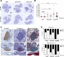
          In this study, we used RNA sequencing analysis to investigate gene-expression profiling of GLO1-depleted breast cancer cells and we validated the regulated expression of selected genes of interest by RT-qPCR. Using in vitro and in vivo assays, we demonstrated the acquisition of a pro-metastatic phenotype related to dicarbonyl stress in MDA-MB-231, MDA-MB-468 and MCF7 breast cancer cellular models. Hyperactivation of MEK/ERK/SMAD1 pathway was evidenced using western blotting upon endogenous MG stress and exogenous MG treatment conditions. MEK and SMAD1 regulation of MG pro-metastatic signature genes in breast cancer cells was demonstrated by RT-qPCR.

          Results

          High-throughput transcriptome profiling of GLO1-depleted breast cancer cells highlighted a pro-metastatic signature that establishes novel connections between MG dicarbonyl stress, extracellular matrix (ECM) remodeling by neoplastic cells and enhanced cell migration. Mechanistically, we showed that these metastasis-related processes are functionally linked to MEK/ERK/SMAD1 cascade activation in breast cancer cells. We showed that sustained MEK/ERK activation in GLO1-depleted cells notably occurred through the down-regulation of the expression of dual specificity phosphatases in MG-stressed breast cancer cells. The use of carnosine and aminoguanidine, two potent MG scavengers, reversed MG stress effects in in vitro and in vivo experimental settings.

          Conclusions

          These results uncover for the first time the key role of MG dicarbonyl stress in the induction of ECM remodeling and the activation of migratory signaling pathways, both in favor of enhanced metastatic dissemination of breast cancer cells. Importantly, the efficient inhibition of mitogen-activated protein kinase (MAPK) signaling using MG scavengers further emphasizes the need to investigate their therapeutic potential across different malignancies.

          Electronic supplementary material

          The online version of this article (10.1186/s13058-018-1095-7) contains supplementary material, which is available to authorized users.

          Related collections

          Most cited references29

          • Record: found
          • Abstract: found
          • Article: not found

          Differential regulation of MAP kinase signalling by dual-specificity protein phosphatases.

          The regulated dephosphorylation of mitogen-activated protein kinases (MAPKs) plays a key role in determining the magnitude and duration of kinase activation and hence the physiological outcome of signalling. In mammalian cells, an important component of this control is mediated by the differential expression and activities of a family of 10 dual-specificity (Thr/Tyr) MAPK phosphatases (MKPs). These enzymes share a common structure in which MAPK substrate recognition is determined by sequences within an amino-terminal non-catalytic domain whereas MAPK binding often leads to a conformational change within the C-terminal catalytic domain resulting in increased enzyme activity. MKPs can either recognize and inactivate a single class of MAP kinase, as in the specific inactivation of extracellular signal regulated kinase (ERK) by the cytoplasmic phosphatase DUSP6/MKP-3 or can regulate more than one MAPK pathway as illustrated by the ability of DUSP1/MKP-1 to dephosphorylate ERK, c-Jun amino-terminal kinase and p38 in the cell nucleus. These properties, coupled with transcriptional regulation of MKP expression in response to stimuli that activate MAPK signalling, suggest a complex negative regulatory network in which individual MAPK activities can be subject to negative feedback control, but also raise the possibility that signalling through multiple MAPK pathways may be integrated at the level of regulation by MKPs.
            Bookmark
            • Record: found
            • Abstract: found
            • Article: not found

            A mechanism of repression of TGFbeta/ Smad signaling by oncogenic Ras.

            TGFbeta can override the proliferative effects of EGF and other Ras-activating mitogens in normal epithelial cells. However, epithelial cells harboring oncogenic Ras mutations often show a loss of TGFbeta antimitogenic responses. Here we report that oncogenic Ras inhibits TGFbeta signaling in mammary and lung epithelial cells by negatively regulating the TGFbeta mediators Smad2 and Smad3. Oncogenically activated Ras inhibits the TGFbeta-induced nuclear accumulation of Smad2 and Smad3 and Smad-dependent transcription. Ras acting via Erk MAP kinases causes phosphorylation of Smad2 and Smad3 at specific sites in the region linking the DNA-binding domain and the transcriptional activation domain. These sites are separate from the TGFbeta receptor phosphorylation sites that activate Smad nuclear translocation. Mutation of these MAP kinase sites in Smad3 yields a Ras-resistant form that can rescue the growth inhibitory response to TGFbeta in Ras-transformed cells. EGF, which is weaker than oncogenic mutations at activating Ras, induces a less extensive phosphorylation and cytoplasmic retention of Smad2 and Smad3. Our results suggest a mechanism for the counterbalanced regulation of Smad2/Smad3 by TGFbeta and Ras signals in normal cells, and for the silencing of antimitogenic TGFbeta functions by hyperactive Ras in cancer cells.
              Bookmark
              • Record: found
              • Abstract: found
              • Article: not found

              Altered metabolite levels in cancer: implications for tumour biology and cancer therapy.

              Altered cell metabolism is a characteristic feature of many cancers. Aside from well-described changes in nutrient consumption and waste excretion, altered cancer cell metabolism also results in changes to intracellular metabolite concentrations. Increased levels of metabolites that result directly from genetic mutations and cancer-associated modifications in protein expression can promote cancer initiation and progression. Changes in the levels of specific metabolites, such as 2-hydroxyglutarate, fumarate, succinate, aspartate and reactive oxygen species, can result in altered cell signalling, enzyme activity and/or metabolic flux. In this Review, we discuss the mechanisms that lead to changes in metabolite concentrations in cancer cells, the consequences of these changes for the cells and how they might be exploited to improve cancer therapy.
                Bookmark

                Author and article information

                Contributors
                +32 4 366 25 57 , a.bellahcene@uliege.be
                Journal
                Breast Cancer Res
                Breast Cancer Res
                Breast Cancer Research : BCR
                BioMed Central (London )
                1465-5411
                1465-542X
                23 January 2019
                23 January 2019
                2019
                : 21
                : 11
                Affiliations
                [1 ]ISNI 0000 0001 0805 7253, GRID grid.4861.b, Metastasis Research Laboratory, GIGA-Cancer, , University of Liège (ULiège), ; Pathology Tour, +4 level, Building 23, Avenue Hippocrate 13, 4000 Liège, Belgium
                [2 ]ISNI 0000 0001 0805 7253, GRID grid.4861.b, Genomics Platform, GIGA, , ULiège, ; Liège, Belgium
                [3 ]ISNI 0000 0001 2069 7798, GRID grid.5342.0, Department of Information Technology, , Ghent University, ; Ghent, Belgium
                [4 ]ISNI 0000 0001 0790 3681, GRID grid.5284.b, Translational Cancer Research Unit, , University of Antwerp, ; Antwerp, Belgium
                [5 ]ISNI 0000 0001 0805 7253, GRID grid.4861.b, Laboratory of Experimental Pathology, GIGA-Cancer, , ULiège, ; Liège, Belgium
                [6 ]ISNI 0000 0001 0805 7253, GRID grid.4861.b, Laboratory of Connective Tissues Biology, GIGA-Cancer, , ULiège, ; Liège, Belgium
                [7 ]ISNI 0000 0001 0481 6099, GRID grid.5012.6, Laboratory for Metabolism and Vascular Medicine, Department of Internal Medicine, , Maastricht University, ; Maastricht, The Netherlands
                [8 ]ISNI 0000 0001 0805 7253, GRID grid.4861.b, Laboratory of Hematology, GIGA-Inflammation, Infection and Immunity, , ULiège, ; Liège, Belgium
                [9 ]ISNI 0000 0004 0624 6108, GRID grid.488845.d, Institut de Recherche en Cancérologie de Montpellier, Inserm U1194, ; Montpellier, France
                Author information
                http://orcid.org/0000-0001-9154-7631
                Article
                1095
                10.1186/s13058-018-1095-7
                6343302
                30674353
                296cb3d9-e8d1-4c6d-8038-feedd04c40ae
                © The Author(s). 2019

                Open AccessThis article is distributed under the terms of the Creative Commons Attribution 4.0 International License ( http://creativecommons.org/licenses/by/4.0/), which permits unrestricted use, distribution, and reproduction in any medium, provided you give appropriate credit to the original author(s) and the source, provide a link to the Creative Commons license, and indicate if changes were made. The Creative Commons Public Domain Dedication waiver ( http://creativecommons.org/publicdomain/zero/1.0/) applies to the data made available in this article, unless otherwise stated.

                History
                : 25 June 2018
                : 27 December 2018
                Funding
                Funded by: FundRef http://dx.doi.org/10.13039/501100002661, Fonds De La Recherche Scientifique - FNRS;
                Funded by: FundRef http://dx.doi.org/10.13039/501100005627, Université de Liège;
                Categories
                Research Article
                Custom metadata
                © The Author(s) 2019

                Oncology & Radiotherapy
                breast cancer,methylglyoxal,smad1,metastasis,carnosine,mapk
                Oncology & Radiotherapy
                breast cancer, methylglyoxal, smad1, metastasis, carnosine, mapk

                Comments

                Comment on this article