24
views
0
recommends
+1 Recommend
0 collections
    0
    shares
      • Record: found
      • Abstract: found
      • Article: found
      Is Open Access

      Double-chambered right ventricle in an adult patient diagnosed by transthoracic echocardiography

      research-article

      Read this article at

      Bookmark
          There is no author summary for this article yet. Authors can add summaries to their articles on ScienceOpen to make them more accessible to a non-specialist audience.

          Abstract

          Background

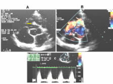
          Double-chambered right ventricle is a rare congenital disease frequently misdiagnosed in the adult patient. An anomalous muscle band divides the right ventricle in two cavities causing variable degree of obstruction. Although echocardiography is considered a useful method for the diagnosis of this pathology in children, it has been recognized the transthoracic scanning limitation in adults.

          Case presentation

          A 29 year-old patient with double-chambered right ventricle presenting mild exercise intolerance referred for follow up of a known ventricular septal defect in whom a complete diagnosis was obtained based only on transthoracic two dimensional echocardiography without the needing of cardiac catheterization.

          Conclusion

          Based on non invasive echocardiographic diagnosis, patient was referred to surgical correction, which was completely successful.

          Related collections

          Most cited references7

          • Record: found
          • Abstract: found
          • Article: not found

          Double-chambered right ventricle presenting in adulthood.

          Double-chambered right ventricle is a form of right ventricular outflow tract obstruction that develops over time, often in patients with an abnormally short distance between the moderator band and pulmonary valve. This lesion typically presents in childhood or adolescence and is often accompanied by a ventricular septal defect. Only a handful of previous cases have been described in which double-chambered right ventricle occurred in adulthood. Since 1992, three patients more than 30 years old (38, 43, and 66 years of age) have presented at our institution with unusual symptoms or a previous incorrect diagnosis. We reviewed the clinical data in these patients. Presenting symptoms included syncope, angina, and severe dyspnea resembling pulmonary hypertension. In 1 patient, disease was categorized as New York Heart Association class IV, and in the other 2 as class III. Coexisting anomalies included a patent foramen ovale or secundum atrial septal defect in 2 patients, a small ventricular septal defect in 1 (with a probable history of ventricular septal defect in another), and mild aortic regurgitation in 1. All patients required urgent or emergent operations, with peak pressures in the proximal right ventricular chamber of 135 to 180 mm Hg and severely depressed left ventricular function in 1 patient. Resection of the anomalous right ventricular muscle bundles was achieved through a right atrial approach in all patients. All patients were alive with improved functional status at follow-up, which was between 15 and 40 months. Right ventricular outflow tract obstruction resulting from a double-chambered right ventricle is rare in adults, but when it does occur it can present with unusual symptoms. When evaluating the patient with signs or symptoms of primary right heart failure, cardiologists should make an effort to image the entire right heart complex. Subcostal echocardiography can facilitate adequate visualization of the right ventricle when it is difficult to distinguish the subpulmonary outflow tract from the parasternal and apical windows.
            Bookmark
            • Record: found
            • Abstract: found
            • Article: not found

            The role of echocardiography in diagnosing double chambered right ventricle in adults.

            To evaluate the utility of echocardiography in diagnosing double chambered right ventricle (DCRV) in adults. A retrospective study. Department of Congenital Heart Diseases and Department of Cardiac Surgery, Institute of Cardiology, Warsaw, Poland. 32 patients, mean age 32.5 years, 21 female, and 11 male. Transthoracic (TTE) and transoesophageal (TOE) echocardiographic examination in patients with suspected DCRV. Direct inspection during surgical treatment of 28 patients diagnosed as having DCRV as an isolated lesion or associated with other pathologies. Echocardiography allowed the final diagnosis of DCRV in 26 patients (81%) out of 32 studied. TTE was diagnostic in 5 (15.6%) whereas TOE was diagnostic in 21 of 21 studied by this technique. Of 6 patients with negative TTE, DCRV was identified by cardiac catheterisation in 3 and directly during surgery in the remaining 3. Of 26 patients diagnosed by echocardiography, the anomalous muscle bundle was discrete in 20 (77%) and diffuse in 6 (23%). In 23 patients (88%) right ventricular outflow obstruction was localised low in the right ventricle, and in the remaining 3 (11.5%), the obstruction was localised high in the right ventricle. Abnormal bundles localised high were discrete, bundles localised in the lower part of right ventricle were discrete in 17 (74%) and diffuse in the remaining 6 (26%). DCRV was an isolated lesion in only 2 patients (6.2%). In all subjects surgical inspection confirmed echocardiographic data. Echocardiography was very useful to diagnose DCRV in adults as well as to identify its anatomic type. Comparing two different approaches, TOE better defines the entire scope of pathology, including estimation of the resultant systolic pressure gradient within the right ventricular cavity.
              Bookmark
              • Record: found
              • Abstract: not found
              • Article: not found

              Difficulty in diagnosing double-chambered right ventricle in adults.

                Bookmark

                Author and article information

                Journal
                Cardiovasc Ultrasound
                Cardiovascular Ultrasound
                BioMed Central (London )
                1476-7120
                2007
                4 January 2007
                : 5
                : 2
                Affiliations
                [1 ]Division of Cardiology, Department of Internal Medicine, University Hospital, Medical School of Ribeirão Preto, University of São Paulo, Ribeirão Preto, SP, Brazil
                Article
                1476-7120-5-2
                10.1186/1476-7120-5-2
                1794404
                17204137
                28d125d7-abd3-4093-b7d4-edc514402eec
                Copyright © 2007 Romano et al; licensee BioMed Central Ltd.

                This is an Open Access article distributed under the terms of the Creative Commons Attribution License ( http://creativecommons.org/licenses/by/2.0), which permits unrestricted use, distribution, and reproduction in any medium, provided the original work is properly cited.

                History
                : 10 November 2006
                : 4 January 2007
                Categories
                Case Report

                Cardiovascular Medicine
                Cardiovascular Medicine

                Comments

                Comment on this article