9
views
0
recommends
+1 Recommend
0 collections
    0
    shares
      • Record: found
      • Abstract: found
      • Article: found
      Is Open Access

      Atlantic salmon skin barrier functions gradually enhance after seawater transfer

      research-article

      Read this article at

      Bookmark
          There is no author summary for this article yet. Authors can add summaries to their articles on ScienceOpen to make them more accessible to a non-specialist audience.

          Abstract

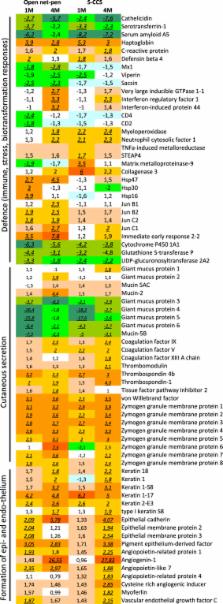
          Atlantic salmon farming operates with high production intensities where skin integrity is recognized as a central factor and indicator for animal health and welfare. In the described trial, the skin development and its immune status in healthy Atlantic salmon reared in two different systems, a traditional open net-pen system and a semi-closed containment system, were investigated. Freshwater smolts were compared to post-smolts after 1 and 4 months in seawater. Growth performance, when adjusted for temperature, was equal between the systems. Skin analyses, including epidermis and dermis, showed that thickness and mucus cell numbers increased in pace with the growth and time post seawater transfer (PST). Gene expression changes suggested similar processes with development of connective tissue, formation of extracellular matrix and augmented cutaneous secretion, changes in mucus protein composition and overall increased immune activity related to gradually enforced protection against pathogens. Results suggest a gradual morphological development in skin with a delayed recovery of immune functions PST. It is possible that Atlantic salmon could experience increased susceptibility to infectious agents and risk of diseases during the first post-smolt period.

          Related collections

          Most cited references35

          • Record: found
          • Abstract: found
          • Article: not found

          Crosstalk between B lymphocytes, microbiota and the intestinal epithelium governs immunity versus metabolism in the gut.

          Using a systems biology approach, we discovered and dissected a three-way interaction between the immune system, the intestinal epithelium and the microbiota. We found that, in the absence of B cells, or of IgA, and in the presence of the microbiota, the intestinal epithelium launches its own protective mechanisms, upregulating interferon-inducible immune response pathways and simultaneously repressing Gata4-related metabolic functions. This shift in intestinal function leads to lipid malabsorption and decreased deposition of body fat. Network analysis revealed the presence of two interconnected epithelial-cell gene networks, one governing lipid metabolism and another regulating immunity, that were inversely expressed. Gene expression patterns in gut biopsies from individuals with common variable immunodeficiency or with HIV infection and intestinal malabsorption were very similar to those of the B cell-deficient mice, providing a possible explanation for a longstanding enigmatic association between immunodeficiency and defective lipid absorption in humans.
            Bookmark
            • Record: found
            • Abstract: found
            • Article: found
            Is Open Access

            Local and systemic gene expression responses of Atlantic salmon (Salmo salar L.) to infection with the salmon louse (Lepeophtheirus salmonis)

            Background The salmon louse (SL) is an ectoparasitic caligid crustacean infecting salmonid fishes in the marine environment. SL represents one of the major challenges for farming of salmonids, and veterinary intervention is necessary to combat infection. This study addressed gene expression responses of Atlantic salmon infected with SL, which may account for its high susceptibility. Results The effects of SL infection on gene expression in Atlantic salmon were studied throughout the infection period from copepodids at 3 days post infection (dpi) to adult lice (33 dpi). Gene expression was analyzed at three developmental stages in damaged and intact skin, spleen, head kidney and liver, using real-time qPCR and a salmonid cDNA microarray (SFA2). Rapid detection of parasites was indicated by the up-regulation of immunoglobulins in the spleen and head kidney and IL-1 receptor type 1, CD4, beta-2-microglobulin, IL-12β, CD8α and arginase 1 in the intact skin of infected fish. Most immune responses decreased at 22 dpi, however, a second activation was observed at 33 dpi. The observed pattern of gene expression in damaged skin suggested the development of inflammation with signs of Th2-like responses. Involvement of T cells in responses to SL was witnessed with up-regulation of CD4, CD8α and programmed death ligand 1. Signs of hyporesponsive immune cells were seen. Cellular stress was prevalent in damaged skin as seen by highly significant up-regulation of heat shock proteins, other chaperones and mitochondrial proteins. Induction of the major components of extracellular matrix, TGF-β and IL-10 was observed only at the adult stage of SL. Taken together with up-regulation of matrix metalloproteinases (MMP), this classifies the wounds afflicted by SL as chronic. Overall, the gene expression changes suggest a combination of chronic stress, impaired healing and immunomodulation. Steady increase of MMP expression in all tissues except liver was a remarkable feature of SL infected fish. Conclusion SL infection in Atlantic salmon is associated with a rapid induction of mixed inflammatory responses, followed by a period of hyporesponsiveness and delayed healing of injuries. Persistent infection may lead to compromised host immunity and tissue self-destruction.
              Bookmark
              • Record: found
              • Abstract: found
              • Article: not found

              Matrix metalloproteinase 9 modulates collagen matrices and wound repair.

              Acute and chronic injuries are characterized by leukocyte infiltration into tissues. Although matrix metalloproteinase 9 (Mmp9) has been implicated in both conditions, its role in wound repair remains unclear. We previously reported a zebrafish chronic inflammation mutant caused by an insertion in the hepatocyte growth factor activator inhibitor gene 1 (hai1; also known as spint1) that is characterized by epithelial extrusions and neutrophil infiltration into the fin. Here, we performed a microarray analysis and found increased inflammatory gene expression in the mutant larvae, including a marked increase in mmp9 expression. Depletion of mmp9 partially rescued the chronic inflammation and epithelial phenotypes, in addition to restoring collagen fiber organization, as detected by second-harmonic generation imaging. Additionally, we found that acute wounding induces epithelial cell mmp9 expression and is associated with a thickening of collagen fibers. Interestingly, depletion of mmp9 impaired this collagen fiber reorganization. Moreover, mmp9 depletion impaired tissue regeneration after tail transection, implicating Mmp9 in acute wound repair. Thus, Mmp9 regulates both acute and chronic tissue damage and plays an essential role in collagen reorganization during wound repair. © 2015. Published by The Company of Biologists Ltd.
                Bookmark

                Author and article information

                Contributors
                christian.karlsen@nofima.no
                Journal
                Sci Rep
                Sci Rep
                Scientific Reports
                Nature Publishing Group UK (London )
                2045-2322
                22 June 2018
                22 June 2018
                2018
                : 8
                : 9510
                Affiliations
                [1 ]ISNI 0000 0004 0451 2652, GRID grid.22736.32, Nofima, Osloveien 1, ; 1430 Aas, Norway
                [2 ]GRID grid.426489.5, UNI Research, Nygårdsgaten 112, ; 5008 Bergen, Norway
                Article
                27818
                10.1038/s41598-018-27818-y
                6015023
                29934588
                283e7ef5-67a0-464e-b2cb-c43661f31e39
                © The Author(s) 2018

                Open Access This article is licensed under a Creative Commons Attribution 4.0 International License, which permits use, sharing, adaptation, distribution and reproduction in any medium or format, as long as you give appropriate credit to the original author(s) and the source, provide a link to the Creative Commons license, and indicate if changes were made. The images or other third party material in this article are included in the article’s Creative Commons license, unless indicated otherwise in a credit line to the material. If material is not included in the article’s Creative Commons license and your intended use is not permitted by statutory regulation or exceeds the permitted use, you will need to obtain permission directly from the copyright holder. To view a copy of this license, visit http://creativecommons.org/licenses/by/4.0/.

                History
                : 14 March 2018
                : 7 June 2018
                Categories
                Article
                Custom metadata
                © The Author(s) 2018

                Uncategorized
                Uncategorized

                Comments

                Comment on this article