24
views
0
recommends
+1 Recommend
0 collections
    0
    shares
      • Record: found
      • Abstract: found
      • Article: found
      Is Open Access

      Quantitative assessment of background parenchymal enhancement in breast MRI predicts response to risk-reducing salpingo-oophorectomy: preliminary evaluation in a cohort of BRCA1/2 mutation carriers

      research-article

      Read this article at

      Bookmark
          There is no author summary for this article yet. Authors can add summaries to their articles on ScienceOpen to make them more accessible to a non-specialist audience.

          Abstract

          Introduction

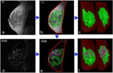
          We present a fully automated method for deriving quantitative measures of background parenchymal enhancement (BPE) from breast dynamic contrast-enhanced magnetic resonance imaging (DCE-MRI) and perform a preliminary evaluation of these measures to assess the effect of risk-reducing salpingo-oophorectomy (RRSO) in a cohort of breast cancer susceptibility gene 1/2 ( BRCA1/2) mutation carriers.

          Methods

          Breast DCE-MRI data from 50 BRCA1/2 carriers were retrospectively analyzed in compliance with the Health Insurance Portability and Accountability Act and with institutional review board approval. Both the absolute (| |) and relative (%) measures of BPE and fibroglandular tissue (FGT) were computed from the MRI scans acquired before and after RRSO. These pre-RRSO and post-RRSO measures were compared using paired Student’s t test. The area under the curve (AUC) of the receiver operating characteristic (ROC) was used to evaluate the performance of relative changes in the BPE and FGT measures in predicting breast cancer that developed in these women after the RRSO surgery.

          Results

          For the 44 women who did not develop breast cancer after RRSO, the absolute volume of BPE and FGT had a significant decrease ( P < 0.05) post-RRSO, whereas for the 6 women who developed breast cancer, there were no significant changes in these measures. Higher values in all BPE and FGT measures were also observed post-RRSO for the women who developed breast cancer, compared with women who did not. Relative changes in BPE percentage were most predictive of women who developed breast cancer after RRSO ( P < 0.05), whereas combining BPE percentage and |FGT| yielded an AUC of 0.80, higher than BPE percentage (AUC = 0.78) or |FGT| (AUC = 0.66) alone (both P > 0.02).

          Conclusions

          Quantitative measures of BPE and FGT are different before and after RRSO, and their relative changes are associated with prediction of developing breast cancer, potentially indicative of women who are more susceptible to develop breast cancer after RRSO in BRCA1/2 mutation carriers.

          Electronic supplementary material

          The online version of this article (doi:10.1186/s13058-015-0577-0) contains supplementary material, which is available to authorized users.

          Related collections

          Most cited references25

          • Record: found
          • Abstract: found
          • Article: not found

          Mutual-information-based registration of medical images: a survey.

          An overview is presented of the medical image processing literature on mutual-information-based registration. The aim of the survey is threefold: an introduction for those new to the field, an overview for those working in the field, and a reference for those searching for literature on a specific application. Methods are classified according to the different aspects of mutual-information-based registration. The main division is in aspects of the methodology and of the application. The part on methodology describes choices made on facets such as preprocessing of images, gray value interpolation, optimization, adaptations to the mutual information measure, and different types of geometrical transformations. The part on applications is a reference of the literature available on different modalities, on interpatient registration and on different anatomical objects. Comparison studies including mutual information are also considered. The paper starts with a description of entropy and mutual information and it closes with a discussion on past achievements and some future challenges.
            Bookmark
            • Record: found
            • Abstract: found
            • Article: not found

            Dynamic breast MR imaging: are signal intensity time course data useful for differential diagnosis of enhancing lesions?

            To assess the relevance of the signal intensity time course for the differential diagnosis of enhancing lesions in dynamic magnetic resonance (MR) imaging of the breast. Two hundred sixty-six breast lesions were examined with a two-dimensional dynamic MR imaging series and subtraction postprocessing. Time-signal intensity curves of the lesions were obtained and classified according to their shapes as type I, which was steady enhancement; type II, plateau of signal intensity; or type III, washout of signal intensity. Enhancement rates and curve types of benign and malignant lesions were compared. There were 101 malignant and 165 benign lesions. The distribution of curve types for breast cancers was type I, 8.9%; type II, 33.6%; and type III, 57.4%. The distribution of curve types for benign lesions was type I, 83.0%; type II, 11.5%; and type III, 5.5%. The distributions proved significantly different (chi 2 = 139.6; P < .001). The diagnostic indices for signal intensity time course were sensitivity, 91%; specificity, 83%; and diagnostic accuracy, 86%. The diagnostic indices for the enhancement rate were sensitivity, 91%; specificity, 37%; and diagnostic accuracy, 58%. The shape of the time-signal intensity curve is an important criterion in differentiating benign and malignant enhancing lesions in dynamic breast MR imaging. A type III time course is a strong indicator of malignancy and is independent of other criteria.
              Bookmark
              • Record: found
              • Abstract: found
              • Article: not found

              American Cancer Society guidelines for breast screening with MRI as an adjunct to mammography.

              New evidence on breast Magnetic Resonance Imaging (MRI) screening has become available since the American Cancer Society (ACS) last issued guidelines for the early detection of breast cancer in 2003. A guideline panel has reviewed this evidence and developed new recommendations for women at different defined levels of risk. Screening MRI is recommended for women with an approximately 20-25% or greater lifetime risk of breast cancer, including women with a strong family history of breast or ovarian cancer and women who were treated for Hodgkin disease. There are several risk subgroups for which the available data are insufficient to recommend for or against screening, including women with a personal history of breast cancer, carcinoma in situ, atypical hyperplasia, and extremely dense breasts on mammography. Diagnostic uses of MRI were not considered to be within the scope of this review.
                Bookmark

                Author and article information

                Contributors
                wus3@upmc.edu
                susan.weinstein@uphs.upenn.edu
                michael.deleo@uphs.upenn.edu
                emily.conant@uphs.upenn.edu
                jinboche@mail.med.upenn.edu
                susan.domchek@uphs.upenn.edu
                despina.kontos@uphs.upenn.edu
                Journal
                Breast Cancer Res
                Breast Cancer Research : BCR
                BioMed Central (London )
                1465-5411
                1465-542X
                19 May 2015
                19 May 2015
                2015
                : 17
                : 1
                : 67
                Affiliations
                [ ]Department of Radiology, Hospital of the University of Pennsylvania, 1 Silverstein Building, 3400 Spruce Street, Philadelphia, PA 19104 USA
                [ ]Department of Biostatistics and Epidemiology, Perelman School of Medicine, University of Pennsylvania, 203 Blockley Hall, 423 Guardian Drive, Philadelphia, PA USA
                [ ]Department of Medicine, Hospital of the University of Pennsylvania, 3400 Civic Center Boulevard, 3 West Pavilion, Philadelphia, PA 19104 USA
                [ ]Present address: Imaging Research Division, Department of Radiology, University of Pittsburgh, 3362 Fifth Avenue, Pittsburgh, PA 15213 USA
                Article
                577
                10.1186/s13058-015-0577-0
                4481125
                25986460
                261c44c2-6b0b-4cd3-ba37-3dd295354d53
                © Wu et al. 2015

                This is an Open Access article distributed under the terms of the Creative Commons Attribution License ( http://creativecommons.org/licenses/by/4.0), which permits unrestricted use, distribution, and reproduction in any medium, provided the original work is properly credited. The Creative Commons Public Domain Dedication waiver ( http://creativecommons.org/publicdomain/zero/1.0/) applies to the data made available in this article, unless otherwise stated.

                History
                : 24 October 2014
                : 11 May 2015
                Categories
                Research Article
                Custom metadata
                © The Author(s) 2015

                Oncology & Radiotherapy
                Oncology & Radiotherapy

                Comments

                Comment on this article