7
views
0
recommends
+1 Recommend
0 collections
    0
    shares
      • Record: found
      • Abstract: found
      • Article: found
      Is Open Access

      Requirements for the differentiation of innate T-bet high memory-phenotype CD4 + T lymphocytes under steady state

      research-article

      Read this article at

      Bookmark
          There is no author summary for this article yet. Authors can add summaries to their articles on ScienceOpen to make them more accessible to a non-specialist audience.

          Abstract

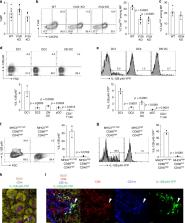
          CD4 + T lymphocytes consist of naïve, antigen-specific memory, and memory-phenotype (MP) cell compartments at homeostasis. We recently showed that MP cells exert innate-like effector function during host defense, but whether MP CD4 + T cells are functionally heterogeneous and, if so, what signals specify the differentiation of MP cell subpopulations under homeostatic conditions is still unclear. Here we characterize MP lymphocytes as consisting of T-bet high, T-bet low, and T-bet subsets, with innate, Th1-like effector activity exclusively associated with T-bet high cells. We further show that the latter population depends on IL-12 produced by CD8α + type 1 dendritic cells (DC1) for its differentiation. Finally, our data demonstrate that this tonic IL-12 production requires TLR-MyD88 signaling independent of foreign agonists, and is further enhanced by CD40-CD40L interactions between DC1 and CD4 + T lymphocytes. We propose that optimal differentiation of T-bet high MP lymphocytes at homeostasis is driven by self-recognition signals at both the DC and Tcell levels.

          Abstract

          CD4 + T cells contain a T-bet high memory-phenotype (MP) population with innate-like functions. Here the authors characterize the requirements for their differentiation at homeostasis and identify a function for IL-12 that is tonically produced by type 1 dendritic cells in an MyD88- and CD40-dependent, but foreign PAMP-independent manner.

          Related collections

          Most cited references46

          • Record: found
          • Abstract: found
          • Article: not found

          Batf3 deficiency reveals a critical role for CD8alpha+ dendritic cells in cytotoxic T cell immunity.

          Although in vitro observations suggest that cross-presentation of antigens is mediated primarily by CD8alpha+ dendritic cells, in vivo analysis has been hampered by the lack of systems that selectively eliminate this cell lineage. We show that deletion of the transcription factor Batf3 ablated development of CD8alpha+ dendritic cells, allowing us to examine their role in immunity in vivo. Dendritic cells from Batf3-/- mice were defective in cross-presentation, and Batf3-/- mice lacked virus-specific CD8+ T cell responses to West Nile virus. Importantly, rejection of highly immunogenic syngeneic tumors was impaired in Batf3-/- mice. These results suggest an important role for CD8alpha+ dendritic cells and cross-presentation in responses to viruses and in tumor rejection.
            Bookmark
            • Record: found
            • Abstract: found
            • Article: not found

            Differential Roles of TLR2 and TLR4 in Recognition of Gram-Negative and Gram-Positive Bacterial Cell Wall Components

            Toll-like receptor (TLR) 2 and TLR4 are implicated in the recognition of various bacterial cell wall components, such as lipopolysaccharide (LPS). To investigate in vivo roles of TLR2, we generated TLR2-deficient mice. In contrast to LPS unresponsiveness in TLR4-deficient mice, TLR2-deficient mice responded to LPS to the same extent as wild-type mice. TLR2-deficient macrophages were hyporesponsive to several Gram-positive bacterial cell walls as well as Staphylococcus aureus peptidoglycan. TLR4-deficient macrophages lacked the response to Gram-positive lipoteichoic acids. These results demonstrate that TLR2 and TLR4 recognize different bacterial cell wall components in vivo and TLR2 plays a major role in Gram-positive bacterial recognition.
              Bookmark
              • Record: found
              • Abstract: found
              • Article: not found

              Recognition of single-stranded RNA viruses by Toll-like receptor 7.

              Viral infection of mammalian host results in the activation of innate immune responses. Toll-like receptors (TLRs) have been shown to mediate the recognition of many types of pathogens, including viruses. The genomes of viruses possess unique characteristics that are not found in mammalian genomes, such as high CpG content and double-stranded RNA. These genomic nucleic acids serve as molecular signatures associated with viral infections. Here we show that TLR7 recognizes the single-stranded RNA viruses, vesicular stomatitis virus and influenza virus. The recognition of these viruses by plasmacytoid dendritic cells and B cells through TLR7 results in their activation of costimulatory molecules and production of cytokines. Moreover, this recognition required intact endocytic pathways. Mice deficient in either the TLR7 or the TLR adaptor protein MyD88 demonstrated reduced responses to in vivo infection with vesicular stomatitis virus. These results demonstrate microbial ligand recognition by TLR7 and provide insights into the pathways used by the innate immune cells in the recognition of viral pathogens.
                Bookmark

                Author and article information

                Contributors
                kawabet@med.tohoku.ac.jp
                asher@niaid.nih.gov
                Journal
                Nat Commun
                Nat Commun
                Nature Communications
                Nature Publishing Group UK (London )
                2041-1723
                6 July 2020
                6 July 2020
                2020
                : 11
                : 3366
                Affiliations
                [1 ]ISNI 0000 0001 2164 9667, GRID grid.419681.3, Immunobiology Section, , Laboratory of Parasitic Diseases, National Institute of Allergy and Infectious Diseases (NIAID), National Institutes of Health (NIH), ; Bethesda, MD 20892 USA
                [2 ]ISNI 0000 0001 2248 6943, GRID grid.69566.3a, Department of Microbiology and Immunology, , Tohoku University Graduate School of Medicine, ; Sendai, 980-8575 Japan
                [3 ]ISNI 0000 0004 1784 4496, GRID grid.410720.0, Academy of Immunology and Microbiology, Institute for Basic Science, ; Pohang, 37666 Republic of Korea
                [4 ]ISNI 0000 0001 0742 4007, GRID grid.49100.3c, Department of Integrative Biosciences and Biotechnology, , Pohang University of Science and Technology, ; Pohang, 37673 Republic of Korea
                [5 ]Molecular and Cellular Immunoregulation Section, Laboratory of Immune System Biology, NIAID, NIH, Bethesda, MD 20892 USA
                [6 ]Cellular Immunology Section, Vaccine Research Center, NIAID, NIH, Bethesda, MD 20892 USA
                [7 ]ISNI 0000 0001 2237 2479, GRID grid.420086.8, Cancer and Inflammation Program, Center for Cancer Research, National Cancer Institute, NIH, ; Bethesda, MD 20892 USA
                Author information
                http://orcid.org/0000-0001-7173-4472
                http://orcid.org/0000-0003-2105-6743
                http://orcid.org/0000-0001-5892-7464
                http://orcid.org/0000-0001-7053-2895
                Article
                17136
                10.1038/s41467-020-17136-1
                7338451
                32632165
                23f93ac8-6eec-4832-95f0-49accb803c7f
                © This is a U.S. government work and not under copyright protection in the U.S.; foreign copyright protection may apply 2020

                Open Access This article is licensed under a Creative Commons Attribution 4.0 International License, which permits use, sharing, adaptation, distribution and reproduction in any medium or format, as long as you give appropriate credit to the original author(s) and the source, provide a link to the Creative Commons license, and indicate if changes were made. The images or other third party material in this article are included in the article’s Creative Commons license, unless indicated otherwise in a credit line to the material. If material is not included in the article’s Creative Commons license and your intended use is not permitted by statutory regulation or exceeds the permitted use, you will need to obtain permission directly from the copyright holder. To view a copy of this license, visit http://creativecommons.org/licenses/by/4.0/.

                History
                : 27 August 2019
                : 11 June 2020
                Funding
                Funded by: FundRef https://doi.org/10.13039/501100001691, MEXT | Japan Society for the Promotion of Science (JSPS);
                Funded by: FundRef https://doi.org/10.13039/100006492, Division of Intramural Research, National Institute of Allergy and Infectious Diseases (Division of Intramural Research of the NIAID);
                Categories
                Article
                Custom metadata
                © The Author(s) 2020

                Uncategorized
                immunological memory,infection,innate immune cells,cd4-positive t cells
                Uncategorized
                immunological memory, infection, innate immune cells, cd4-positive t cells

                Comments

                Comment on this article