8
views
0
recommends
+1 Recommend
0 collections
    0
    shares
      • Record: found
      • Abstract: found
      • Article: found
      Is Open Access

      CT whole lung radiomic nomogram: a potential biomarker for lung function evaluation and identification of COPD

      research-article

      Read this article at

      Bookmark
          There is no author summary for this article yet. Authors can add summaries to their articles on ScienceOpen to make them more accessible to a non-specialist audience.

          Abstract

          Background

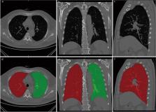
          Computed tomography (CT) plays a great role in characterizing and quantifying changes in lung structure and function of chronic obstructive pulmonary disease (COPD). This study aimed to explore the performance of CT-based whole lung radiomic in discriminating COPD patients and non-COPD patients.

          Methods

          This retrospective study was performed on 2785 patients who underwent pulmonary function examination in 5 hospitals and were divided into non-COPD group and COPD group. The radiomic features of the whole lung volume were extracted. Least absolute shrinkage and selection operator (LASSO) logistic regression was applied for feature selection and radiomic signature construction. A radiomic nomogram was established by combining the radiomic score and clinical factors. Receiver operating characteristic (ROC) curve analysis and decision curve analysis (DCA) were used to evaluate the predictive performance of the radiomic nomogram in the training, internal validation, and independent external validation cohorts.

          Results

          Eighteen radiomic features were collected from the whole lung volume to construct a radiomic model. The area under the curve (AUC) of the radiomic model in the training, internal, and independent external validation cohorts were 0.888 [95% confidence interval (CI) 0.869–0.906], 0.874 (95%CI 0.844–0.904) and 0.846 (95%CI 0.822–0.870), respectively. All were higher than the clinical model (AUC were 0.732, 0.714, and 0.777, respectively, P < 0.001). DCA demonstrated that the nomogram constructed by combining radiomic score, age, sex, height, and smoking status was superior to the clinical factor model.

          Conclusions

          The intuitive nomogram constructed by CT-based whole-lung radiomic has shown good performance and high accuracy in identifying COPD in this multicenter study.

          Supplementary Information

          The online version contains supplementary material available at 10.1186/s40779-024-00516-9.

          Related collections

          Most cited references31

          • Record: found
          • Abstract: found
          • Article: not found

          Prevalence and risk factors of chronic obstructive pulmonary disease in China (the China Pulmonary Health [CPH] study): a national cross-sectional study

          Although exposure to cigarette smoking and air pollution is common, the current prevalence of chronic obstructive pulmonary disease (COPD) is unknown in the Chinese adult population. We conducted the China Pulmonary Health (CPH) study to assess the prevalence and risk factors of COPD in China.
            Bookmark
            • Record: found
            • Abstract: not found
            • Article: not found

            Introduction to Radiomics

              Bookmark
              • Record: found
              • Abstract: not found
              • Article: not found

              A review of feature selection methods in medical applications

                Bookmark

                Author and article information

                Contributors
                fanli0930@163.com , czyxfl@smmu.edu.cn
                Journal
                Mil Med Res
                Mil Med Res
                Military Medical Research
                BioMed Central (London )
                2095-7467
                2054-9369
                20 February 2024
                20 February 2024
                2024
                : 11
                : 14
                Affiliations
                [1 ]GRID grid.73113.37, ISNI 0000 0004 0369 1660, Department of Radiology, , the Second Affiliated Hospital of Naval Medical University, ; Shanghai, 200003 China
                [2 ]School of Medical Imaging, Shandong Second Medical University, Weifang, 261053 Shandong China
                [3 ]GRID grid.412793.a, ISNI 0000 0004 1799 5032, Department of Radiology, School of Medicine, , Tongji Hospital, Tongji University, ; Shanghai, 200065 China
                [4 ]GRID grid.506977.a, ISNI 0000 0004 1757 7957, Department of Radiology, , Zhejiang Province People’s Hospital, Affiliated People’s Hospital of Hangzhou Medical College, ; Hangzhou, 310014 China
                [5 ]Department of Radiology, Sir Run Run Shaw Hospital, ( https://ror.org/00ka6rp58) Zhejiang, 310018 China
                [6 ]GRID grid.415002.2, ISNI 0000 0004 1757 8108, Jiangxi Provincial People’s Hospital, , the First Affiliated Hospital of Nanchang Medical College, ; Nanchang, 330006 China
                [7 ]College of Health Sciences and Engineering, University of Shanghai for Science and Technology, ( https://ror.org/00ay9v204) Shanghai, 200093 China
                [8 ]Department of Radiology, the Second People’s Hospital of Deyang, ( https://ror.org/042g3qa69) Deyang, 618000 Sichuan China
                [9 ]Department of Radiation Oncology, Shanghai Jiao Tong University Affiliated Sixth People’s Hospital, ( https://ror.org/0220qvk04) Shanghai, 200233 China
                Article
                516
                10.1186/s40779-024-00516-9
                10877876
                38374260
                23f2065d-5ff7-4a73-8c3b-e616bc3a5c99
                © The Author(s) 2024

                Open Access This article is licensed under a Creative Commons Attribution 4.0 International License, which permits use, sharing, adaptation, distribution and reproduction in any medium or format, as long as you give appropriate credit to the original author(s) and the source, provide a link to the Creative Commons licence, and indicate if changes were made. The images or other third party material in this article are included in the article's Creative Commons licence, unless indicated otherwise in a credit line to the material. If material is not included in the article's Creative Commons licence and your intended use is not permitted by statutory regulation or exceeds the permitted use, you will need to obtain permission directly from the copyright holder. To view a copy of this licence, visit http://creativecommons.org/licenses/by/4.0/. The Creative Commons Public Domain Dedication waiver ( http://creativecommons.org/publicdomain/zero/1.0/) applies to the data made available in this article, unless otherwise stated in a credit line to the data.

                History
                : 4 July 2023
                : 22 January 2024
                Funding
                Funded by: FundRef http://dx.doi.org/10.13039/501100001809, National Natural Science Foundation of China;
                Award ID: 82171926
                Award ID: 81930049
                Award ID: 82202140
                Award Recipient :
                Funded by: National Key R&D Program of China
                Award ID: 2022YFC2010002
                Award ID: 2022YFC2010000
                Award ID: 2022YFC2010005
                Award Recipient :
                Funded by: Medical imaging database construction program of National Health Comission
                Award ID: YXFSC2022JJSJ002
                Award Recipient :
                Funded by: clinical Innovative Project of Shanghai Changzheng Hospital
                Award ID: 2020YLCYJ-Y24
                Award Recipient :
                Funded by: program of Science and Technology Commission of Shanghai Municipality
                Award ID: 21DZ2202600
                Award Recipient :
                Funded by: Shanghai Sailing Program
                Award ID: 20YF1449000
                Award Recipient :
                Categories
                Research
                Custom metadata
                © People´s Military Medical Press 2024

                chronic obstructive pulmonary disease (copd),computed tomography (ct),radiomic

                Comments

                Comment on this article