31
views
0
recommends
+1 Recommend
0 collections
    0
    shares
      • Record: found
      • Abstract: found
      • Article: not found

      A structural analysis of M protein in coronavirus assembly and morphology

      research-article

      Read this article at

      ScienceOpenPublisherPMC
      Bookmark
          There is no author summary for this article yet. Authors can add summaries to their articles on ScienceOpen to make them more accessible to a non-specialist audience.

          Abstract

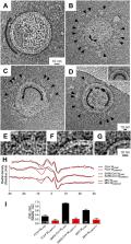
          The M protein of coronavirus plays a central role in virus assembly, turning cellular membranes into workshops where virus and host factors come together to make new virus particles. We investigated how M structure and organization is related to virus shape and size using cryo-electron microscopy, tomography and statistical analysis. We present evidence that suggests M can adopt two conformations and that membrane curvature is regulated by one M conformer. Elongated M protein is associated with rigidity, clusters of spikes and a relatively narrow range of membrane curvature. In contrast, compact M protein is associated with flexibility and low spike density. Analysis of several types of virus-like particles and virions revealed that S protein, N protein and genomic RNA each help to regulate virion size and variation, presumably through interactions with M. These findings provide insight into how M protein functions to promote virus assembly.

          Related collections

          Most cited references40

          • Record: found
          • Abstract: found
          • Article: not found

          Supramolecular architecture of severe acute respiratory syndrome coronavirus revealed by electron cryomicroscopy.

          Coronavirus particles are enveloped and pleomorphic and are thus refractory to crystallization and symmetry-assisted reconstruction. A novel methodology of single-particle image analysis was applied to selected virus features to obtain a detailed model of the oligomeric state and spatial relationships among viral structural proteins. Two-dimensional images of the S, M, and N structural proteins of severe acute respiratory syndrome coronavirus and two other coronaviruses were refined to a resolution of approximately 4 nm. Proteins near the viral membrane were arranged in overlapping lattices surrounding a disordered core. Trimeric glycoprotein spikes were in register with four underlying ribonucleoprotein densities. However, the spikes were dispensable for ribonucleoprotein lattice formation. The ribonucleoprotein particles displayed coiled shapes when released from the viral membrane. Our results contribute to the understanding of the assembly pathway used by coronaviruses and other pleomorphic viruses and provide the first detailed view of coronavirus ultrastructure.
            Bookmark
            • Record: found
            • Abstract: found
            • Article: not found

            Influenza virus pleiomorphy characterized by cryoelectron tomography.

            Influenza virus remains a global health threat, with millions of infections annually and the impending threat that a strain of avian influenza may develop into a human pandemic. Despite its importance as a pathogen, little is known about the virus structure, in part because of its intrinsic structural variability (pleiomorphy): the primary distinction is between spherical and elongated particles, but both vary in size. Pleiomorphy has thwarted structural analysis by image reconstruction of electron micrographs based on averaging many identical particles. In this study, we used cryoelectron tomography to visualize the 3D structures of 110 individual virions of the X-31 (H3N2) strain of influenza A. The tomograms distinguish two kinds of glycoprotein spikes [hemagglutinin (HA) and neuraminidase (NA)] in the viral envelope, resolve the matrix protein layer lining the envelope, and depict internal configurations of ribonucleoprotein (RNP) complexes. They also reveal the stems that link the glycoprotein ectodomains to the membrane and interactions among the glycoproteins, the matrix, and the RNPs that presumably control the budding of nascent virions from host cells. Five classes of virions, four spherical and one elongated, are distinguished by features of their matrix layer and RNP organization. Some virions have substantial gaps in their matrix layer ("molecular fontanels"), and others appear to lack a matrix layer entirely, suggesting the existence of an alternative budding pathway in which matrix protein is minimally involved.
              Bookmark
              • Record: found
              • Abstract: found
              • Article: not found

              Coronavirus M proteins accumulate in the Golgi complex beyond the site of virion budding.

              The prevailing hypothesis is that the intracellular site of budding of coronaviruses is determined by the localization of its membrane protein M (previously called E1). We tested this by analyzing the site of budding of four different coronaviruses in relation to the intracellular localization of their M proteins. Mouse hepatitis virus (MHV) and infectious bronchitis virus (IBV) grown in Sac(-) cells, and feline infectious peritonitis virus (FIPV) and transmissible gastroenteritis virus (TGEV) grown in CrFK cells, all budded exclusively into smooth-walled, tubulovesicular membranes located intermediately between the rough endoplasmic reticulum and Golgi complex, identical to the so-called budding compartment previously identified for MHV. Indirect immunofluorescence staining of the infected cells showed that all four M proteins accumulated in a perinuclear region. Immunogold microscopy localized MHV M and IBV M in the budding compartment; in addition, a dense labeling in the Golgi complex occurred, MHV M predominantly in trans-Golgi cisternae and trans-Golgi reticulum and IBV M mainly in the cis and medial Golgi cisternae. The corresponding M proteins of the four viruses, when independently expressed in a recombinant vaccinia virus system, also accumulated in the perinuclear area. Quantitative pulse-chase analysis of metabolically labeled cells showed that in each case the majority of the M glycoproteins carried oligosaccharide side chains with Golgi-specific modifications within 4 h after synthesis. Immunoelectron microscopy localized recombinant MHV M and IBV M to the same membranes as the respective proteins in coronavirus-infected cells, with the same cis-trans distribution over the Golgi complex. Our results demonstrate that some of the M proteins of the four viruses are transported beyond the budding compartment and are differentially retained by intrinsic retention signals; in addition to M, other viral and/or cellular factors are probably required to determine the site of budding.
                Bookmark

                Author and article information

                Contributors
                Journal
                J Struct Biol
                J. Struct. Biol
                Journal of Structural Biology
                Elsevier Inc.
                1047-8477
                1095-8657
                3 December 2010
                April 2011
                3 December 2010
                : 174
                : 1
                : 11-22
                Affiliations
                [a ]School of Biological Sciences, University of Reading, RG6 6AJ, UK
                [b ]School of Mathematical and Physical Sciences, University of Reading, RG6 6AJ, UK
                [c ]The Scripps Research Institute, La Jolla, CA, USA
                [d ]Bio-Nanotechnology Laboratory, Department of Neuroscience and Pharmacology, University of Copenhagen, Denmark
                [e ]Nano-Science Center, University of Copenhagen, Denmark
                [f ]Lundbeck Foundation Center for Biomembranes in Nanomedicine, University of Copenhagen, Denmark
                [g ]The University of California, Irvine, CA, USA
                [h ]The University of Texas, Medical Branch, Galveston, TX, USA
                [i ]Medical Research Council Virology Unit, Glasgow, UK
                [j ]University of Toledo, OH, USA
                [k ]University of Bristol, BS8 1TH, UK
                [l ]University of Vermont, Burlington, VT, USA
                Author notes
                [* ]Corresponding author. Fax: +44 118 378 6537. b.w.neuman@ 123456reading.ac.uk
                Article
                S1047-8477(10)00358-8
                10.1016/j.jsb.2010.11.021
                4486061
                21130884
                22c7a25f-619c-41df-a04b-bffa18b81fae
                Copyright © 2010 Elsevier Inc. All rights reserved.

                Since January 2020 Elsevier has created a COVID-19 resource centre with free information in English and Mandarin on the novel coronavirus COVID-19. The COVID-19 resource centre is hosted on Elsevier Connect, the company's public news and information website. Elsevier hereby grants permission to make all its COVID-19-related research that is available on the COVID-19 resource centre - including this research content - immediately available in PubMed Central and other publicly funded repositories, such as the WHO COVID database with rights for unrestricted research re-use and analyses in any form or by any means with acknowledgement of the original source. These permissions are granted for free by Elsevier for as long as the COVID-19 resource centre remains active.

                History
                : 5 July 2010
                : 23 November 2010
                : 26 November 2010
                Categories
                Article

                Biophysics
                cryo-electron microscopy,cryo-electron tomography,pleomorphic virus structure,coronavirus,viral matrix protein

                Comments

                Comment on this article