14
views
0
recommends
+1 Recommend
1 collections
    0
    shares
      • Record: found
      • Abstract: found
      • Article: found
      Is Open Access

      Incidence of reactive hyperplastic lesions in the oral cavity: a 10 year retrospective study in Santa Catarina, Brazil Translated title: Incidência de lesões hiperplásicas reativas na cavidade bucal: estudo retrospectivo de 10 anos em Santa Catarina, Brasil

      research-article

      Read this article at

      ScienceOpenPublisherPMC
      Bookmark
          There is no author summary for this article yet. Authors can add summaries to their articles on ScienceOpen to make them more accessible to a non-specialist audience.

          Abstract

          Introduction

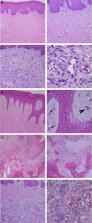
          Reactive hyperplastic lesions develop in response to a chronic injury simulating an exuberant tissue repair response. They represent some of the most common oral lesions including inflammatory fibrous hyperplasia, oral pyogenic granuloma, giant cell fibroma, peripheral ossifying fibroma, and peripheral giant cell lesions.

          Objective

          The incidence of those lesions was investigated in an oral pathology service, and the clinical characteristics, associated etiological factors, concordance between the clinical and histopathological diagnostic was determined.

          Methods

          A total of 2400 patient records were screened from 2006 to 2016. Clinical features were recorded from biopsy reports and patients’ files.

          Results

          A total of 534 cases of reactive hyperplastic lesions were retrieved and retrospectively studied, representing 22.25% of all diagnoses. The most frequent lesion was inflammatory fibrous hyperplasia (72.09%), followed by oral pyogenic granuloma (11.79%), giant cell fibroma (7.30%), peripheral ossifying fibroma (5.24%), and peripheral giant cell lesions (3.55%). Females were predominantly affected (74.19%), the gingiva and alveolar ridge were the predominant anatomical site (32.89%), and chronic traumatism was presented as the main etiological factor. The age widely ranges from the 1st decade of life to the 7th. Clinically, the reactive hyperplastic lesions consisted of small lesions (0.5–2 cm) and shared a strong likeness in color to the oral mucosa. The concordance between the clinical and histopathological diagnostic was high (82.5%).

          Conclusion

          Reactive hyperplastic lesions had a high incidence among oral pathologies. The understanding of their clinical features helps to achieve a clearer clinical and etiological diagnosis, and the knowledge of factors related to their development. This may contribute to adequate treatment and positive prognosis.

          Resumo

          Introdução

          As lesões hiperplásicas reativas se desenvolvem em resposta a uma lesão crônica que estimula uma resposta acentuada de reparo tecidual. Elas representam uma das lesões orais mais comuns, inclusive a hiperplasia fibrosa inflamatória, granuloma piogênico oral, fibroma de células gigantes, fibroma periférico ossificante e lesão periférica de células gigantes.

          Objetivo

          A incidência dessas lesões foi investigada em um serviço de patologia bucal e as características clínicas, os fatores etiológicos associados e a concordância entre os diagnósticos clínico e histopatológico foram determinados.

          Método

          Foram selecionados 2.400 registros de pacientes entre 2006 e 2016. As características clínicas foram registradas a partir de laudos de biópsia e dos prontuários dos pacientes.

          Resultados

          Um total de 534 casos de lesões hiperplásicas reativas foram recuperados e retrospectivamente estudados, representando 22,25% de todos os diagnósticos. A lesão mais frequente foi hiperplasia fibrosa inflamatória (72,09%), seguida por granuloma piogênico oral (11,79%), fibroma de células gigantes, (7,30%), fibroma periférico ossificante (5,24%) e lesão periférica de células gigantes (3,55%). O sexo feminino foi predominante (74,19%), a gengiva e a crista alveolar foram o local anatômico predominante (32,89%) e o traumatismo crônico foi demonstrado como o principal fator etiológico. A idade variou desde a 1ª década de vida até a 7ª. Clinicamente, as LHR consistiram em pequenas lesões (0,5 a 2 cm) que apresentaram uma forte semelhança de cor com a mucosa oral. A concordância entre o diagnóstico clínico e histopatológico foi alta (82,5%).

          Conclusão

          As lesões hiperplásicas reativas apresentaram alta incidência entre as patologias bucais. A compreensão das características clínicas ajuda na realização de um diagnóstico clínico e etiológico mais claro, bem como determinar os fatores relacionados ao seu desenvolvimento. Dessa forma contribui para um tratamento adequado e um prognóstico positivo.

          Related collections

          Most cited references31

          • Record: found
          • Abstract: found
          • Article: found
          Is Open Access

          Oral pyogenic granuloma: Various concepts of etiopathogenesis

          Pyogenic granuloma or granuloma pyogenicum is a well-known oral lesion. The name pyogenic granuloma is a misnomer since the condition is not associated with pus and does not represent a granuloma histologically. Pyogenic granuloma of the oral cavity is known to involve the gingiva commonly. Extragingivally, it can occur on the lips, tongue, buccal mucosa, palate, and the like. A history of trauma is common in such sites. The etiology of the lesion is not known, though it was originally believed to be a botryomycotic infection. It is theorized that pyogenic granuloma possibly originates as a response of tissues to minor trauma and/or chronic irritation, thus opening a pathway for invasion of nonspecific microorganisms, although microorganisms are seldom demonstrated within the lesion. Pathogenesis of pyogenic granuloma is still debatable. Medline and PubMed databases were searched under the following key terms: Pathogenesis of oral pyogenic granuloma, pyogenic granuloma, and oral pyogenic granuloma. This search was limited to articles on human/animal studies which were published in English language. After reviewing the searched articles, the relevant articles were selected for the present review. Through this article, we have tried to summarize and present all the concepts of pathogenesis related to this most common and most mysterious oral lesion.
            Bookmark
            • Record: found
            • Abstract: found
            • Article: not found

            Reactive lesions of the gingiva. A clinicopathological study of 741 cases.

            A series of 741 consecutive cases of localized hyperplastic lesions of the gingiva were studied. The lesions were reclassified into four groups: pyogenic granuloma, peripheral giant cell granuloma, fibrous hyperplasia and peripheral fibroma with calcification. This study indicates that there are some differences between these groups in age and sex distribution as well as in location and size of the lesion. Fibrous hyperplasia was the most common type, followed in descending order by pyogenic granuloma, peripheral fibroma with calcification and peripheral giant cell granuloma. The peripheral giant cell granuloma showed no sex predilection while fibrous hyperplasia, pyogenic granuloma and peripheral fibroma with calcification were more common in females. Pyogenic granuloma and peripheral fibroma with calcification occur in younger patients more often than fibrous hyperplasia, and thus may represent a stage in the development of fibrous hyperplasia.
              Bookmark
              • Record: found
              • Abstract: found
              • Article: not found

              Common benign oral soft tissue masses.

              This article reviews some of the more common benign oral soft tissue masses with emphasis on their etiology, epidemiology, clinical presentation, histopathology, and treatment. These lesions include traumatic fibroma, mucocele, warts/papilloma, pyogenic granuloma, peripheral giant cell granuloma, generalized gingival hyperplasia, gingival fibromatosis, lateral periodontal cyst, lipoma, and denture-induced hyperplasia.
                Bookmark

                Author and article information

                Contributors
                Journal
                Braz J Otorhinolaryngol
                Braz J Otorhinolaryngol
                Brazilian Journal of Otorhinolaryngology
                Elsevier
                1808-8694
                1808-8686
                17 April 2018
                Jul-Aug 2019
                17 April 2018
                : 85
                : 4
                : 399-407
                Affiliations
                [a ]Universidade Federal de Santa Catarina (UFSC), Programa de Pós-Graduação em Odontologia, Florianópolis, SC, Brazil
                [b ]Universidade Federal de Santa Catarina (UFSC), Faculdade de Odontologia, Florianópolis, SC, Brazil
                [c ]Universidade Federal de Santa Catarina (UFSC), Departamento de Patologia, Florianópolis, SC, Brazil
                Author notes
                [* ]Corresponding author. kamileldutra@ 123456gmail.com
                Article
                S1808-8694(18)30095-8
                10.1016/j.bjorl.2018.03.006
                9443062
                29705120
                21c34b11-4372-4b8f-b926-3d92cac95a1f
                © 2018 Associação Brasileira de Otorrinolaringologia e Cirurgia Cérvico-Facial. Published by Elsevier Editora Ltda.

                This is an open access article under the CC BY license (http://creativecommons.org/licenses/by/4.0/).

                History
                : 17 August 2017
                : 15 March 2018
                Categories
                Original Article

                giant cell granuloma,hyperplasia,oral cavity,pyogenic granuloma,reactive hyperplastic lesions,granuloma de células gigantes,hiperplasia,cavidade oral,granuloma piogênico,lesões hiperplásicas reativas

                Comments

                Comment on this article

                scite_

                Similar content69

                Cited by12

                Most referenced authors242