- Record: found
- Abstract: found
- Article: not found
De novo design of a fluorescence-activating β-barrel
research-article
Jiayi Dou
1
,
2 ,
Anastassia A. Vorobieva
1
,
2 ,
William Sheffler
1
,
2 ,
Lindsey A. Doyle
3 ,
Hahnbeom Park
1
,
2 ,
Matthew J. Bick
1
,
2 ,
Binchen Mao
1 ,
Glenna W. Foight
4 ,
Min Yen Lee
4 ,
Lauren A. Gagnon
4 ,
Lauren Carter
1
,
2 ,
Banumathi Sankaran
5 ,
Sergey Ovchinnikov
1
,
2 ,
Enrique Marcos
1
,
2 ,
Po-Ssu Huang
1
,
2 ,
Joshua C. Vaughan
4 ,
Barry L. Stoddard
3 ,
David Baker
1
,
2
,
6
,
#
12 September 2018
Read this article at

There is no author summary for this article yet. Authors can add summaries to their articles on ScienceOpen to make them more accessible to a non-specialist audience.
Abstract
The regular arrangements of β-strands around a central axis in β-barrels and of α-helices
in coiled coils contrasts with the irregular tertiary structures of most globular
proteins, and have fascinated structural biologists since they were first discovered.
Simple parametric models have been used to design a wide range of α-helical coiled
coil structures, but to date there has been no success with β-barrels. Here we first
show that accurate de novo design of β-barrels requires considerable symmetry breaking
to achieve continuous hydrogen bond connectivity and eliminate backbone strain. We
then build ensembles of β-barrel backbone structures with cavity shapes matched to
the fluorogenic compound DFHBI, and use a hierarchical grid-based search method to
simultaneously optimize the rigid body placement of DFHBI in these cavities and the
identities of the surrounding amino acids for high shape and chemically complementary
binding. The designs have high structural accuracy and bind and fluorescently activate
DFHBI in vitro and in E. coli, yeast and mammalian cells. This de novo design of small
molecule binding activity, using backbones custom built to bind the ligand, sets the
stage for design of increasingly sophisticated ligand binding proteins, sensors, and
catalysts not limited by the backbone geometries available in known protein structures.
There have been considerable recent advances in designing protein folds from scratch
1,2
, as well as redesigning already existing native scaffolds to bind small molecules
3–5
, but two outstanding unsolved challenges remain. The first is the de novo design
of all-β proteins, which is complicated by the tendency of β-strands and sheets to
associate intermolecularly to form amyloid like structures if their register is not
perfectly controlled
6
. The second is the design of protein backbones customized to bind small molecules
of interest, which requires precise control over both backbone and sidechain geometry
5
, as well as balancing the often opposing requirements of protein folding and function
7
. Success in developing such methods would reduce the longstanding dependency on natural
proteins by enabling protein engineers to craft new proteins optimized to bind chosen
small-molecule targets, and lay a foundation for de novo design of proteins customized
to catalyze specific chemical reactions.
Principles for designing β-barrels
β-barrels are single β-sheets that twist to form a closed structure in which the first
strand is hydrogen bonded to the last
8
. Anti-parallel β-barrels are excellent scaffolds for ligand binding, as the base
of the barrel can accommodate a hydrophobic core to provide overall stability, and
the top of the barrel can provide a recessed cavity for ligand binding
9
, often flanked by loops that can contribute further binding affinity and selectivity
10
. However β-sheet topologies are notoriously difficult to design from scratch, with
no reported success to date, although several descriptive parametric models of β-barrels
have been proposed
11–13
. We first set out to address this challenge by parametrically generating regular
arrangements of 8 anti-parallel β-strands using the equations for an elliptic hyperboloid
of revolution (adapted from
14
, Extended Data Fig. 1a). β-barrels are characterized by their shear number ‘S’ —
the total shift in strand registry between the first and last strand — which determines
the hydrophobic packing arrangement and the diameter of the barrel (Supplementary
Methods)
15,16
. We selected a shear number of S=10 because it is difficult to achieve good core
packing for S=8 (the barrel has a smaller diameter and the Cα-Cβ vectors point directly
at each other), and S=12 results in a cavity too large to fill with sidechains (Extended
Data Fig. 1b–d). We generated ensembles of hyperboloids by sampling the elliptical
parameters and the tilt of the generating lines with respect to the central axis around
ideal values computed for S=10, and then placed Cαs on the hyperboloid surface (Fig.
1a; Supplementary Methods). As found in earlier simulation work
17
, backbones generated with constant angles between strands could not achieve perfectly
regular hydrogen bonding. To resolve this problem, we introduced force-field guided
variation in local twist by gradient based minimization. We selected the backbones
with the most extensive inter-strand hydrogen bonding, connected the strands with
short loops and carried out combinatorial sequence optimization to obtain low energy
sequences. Synthetic genes encoding 41 such designs were produced and the proteins
expressed in E. coli. Almost all were found to be insoluble or oligomeric; none of
this first set of 41 designs were monomeric with an all-β circular dichroism spectrum
(Supplementary Table 2).
In considering the possible reasons for the failure of the initial designs, we noted
that many of the backbone hydrogen bond interactions on the top and bottom of the
barrels were distorted or broken (Extended Data Fig. 1e,f). To investigate the origins
of this distortion, we experimented with three alternative approaches to generating
uniform β-barrel backbones lacking loops and with valine at every position as a place-holder
(Supplementary Methods). In all cases, we observed breaking of hydrogen bond interactions
following structure minimization with Rosetta relaxation protocol (Extended Data Fig.
2a), suggesting there is strain inherent to the closing of the curved β-sheet on itself.
To identify the origin of this strain, we repeated the relaxation after imposing strong
constraints on the hydrogen bond interactions to prevent them from breaking. As illustrated
in Fig. 1c, the strain manifested in two places. First, steric clashes build up along
strips of side-chains in the directions of the hydrogen bonds, perpendicularly to
the direction of the β-strands (“Cβ-strips”, Fig.1c). Second, a number of residues
acquired unfavorable left handed twist (Extended Data Fig. 2b,c; the chirality of
the peptide backbone favors right handed twist). To reduce the strain arising from
steric clashes between Cβ atoms, and from the local left handed twist, we replaced
the central valine residue of each Cβ-strip with a glycine (which are normally disfavored
in β-sheets
18
). The achiral glycine can have a left-hand twist without disrupting the β-sheet hydrogen
bond pattern
15,19
and lacks a Cβ atom, reducing the steric clashes within Cβ-strips (Fig. 1c, middle).
The backbones of most of these glycine residues shifted to the positive Φ torsion
bin after minimization to form torsional irregularities in the β-sheet (“glycine kinks”
15
, Extended Data Fig. 2d–e).
Based on these observations, we hypothesized that large local deviations in ideal
β-strand twist are necessary to maintain continuous hydrogen bond interactions between
strands in a closed β-barrel, and hence that a parametric approach assuming uniform
geometry was not well suited to building such structures. Therefore, we chose to build
β-barrel backbones starting from a 2D map specifying the peptide bonds, the backbone
torsion angle bins
20
, and the backbone hydrogen bonds (Fig. 1b). In contrast to parametric backbone design,
which may be viewed as “3D to 2D” approach as a 3D surface is generated and then populated
with residues, this alternative strategy proceeds from 2D to 3D and can readily incorporate
local torsional deviation. We generated 3D protein backbones using Rosetta Monte Carlo
structure generation calculations starting from an extended peptide chain
21
, guided by torsional and distance constraints from the 2D map.
We found that we could control the volume and the 3D shape of the β-barrel cavity
by altering the placement of glycine kinks in the 2D map. Such kinks dramatically
increase local β-sheet curvature, forming corners in an otherwise roughly circular
cross-section (Extended Data Fig. 2f,g). We chose to design a square barrel shape
and created four corners in the β-sheet by placing five glycine kinks to un-strain
the five Cβ-strips and one glycine kink to correct the twist of the longest hairpin
(Fig. 1d, Supplementary Methods & Extended Data Fig. 3a). With this choice, the resulting
3D backbones have a large interior volume suitable for a ligand-binding cavity. When
such backbones were built with canonical type I’ β-turns connecting each β-hairpin,
we observed steric strain at the extremities of the Cβ-strips (Fig. 1c, bottom) and
disruption of hydrogen bond interactions following structure relaxation (Extended
Data Fig. 3e). This likely arises because the considerable curvature at the glycine
kinks requires that the β-hairpins paired with it (dashed vertical line in Extended
Data Fig. 3b) must have greater right handed twist than can be achieved with canonical
β-hairpins. We reasoned that accentuated right-handed twist could be achieved by incorporating
β-bulges — disruptions of the regular hydrogen bonding pattern of a β-sheet
2,22,23
. Indeed, we found that strategic placement of β-bulges on the bottom of the barrel
(defined as the side of the N- and C- termini) and bulge-containing β-turns
22
on the top of the barrel eliminated steric strain and stabilized the hydrogen bonds
between the β-strand residues flanking the turns (Extended Data Fig. 3e,f). To tie
together the bottom of the barrel, we introduced a “tryptophan corner”
24,25
by placing a short 3–10 helix followed by a glycine kink and a Trp at the beginning
of the barrel, and an interacting Arg at the C-terminus (Extended Data Fig. 3g–j).
500 backbones were generated from the 2D map incorporating the above features (see
Methods), and Rosetta flexible backbone sequence design calculations were carried
out to identify low energy sequences for each backbone. Four designs with low energy
and backbone hydrogen bonding throughout the barrel were selected for experimental
characterization (Extended Data Fig. 4a). The sequences of these designs are not related
to those of known native proteins with BLAST E-values > 0.1, and fold into the designed
structure in silico (Fig. 2a). Synthetic genes encoding the designs were expressed
in E. coli. Three of the designs were expressed in the soluble fraction and purified;
two had characteristic β-sheet far-UV circular dichroism (CD) signal (Fig. 2; Extended
Data Fig. 4b). Size-exclusion chromatography (SEC) coupled with multi-angle light
scattering (MALS) showed that one was a stable monomer (BB1) and the other (BB2) a
soluble tetramer (Extended Data Fig. 4c).
BB1 exhibited a strong near-UV signature suggesting an organized tertiary structure
(Fig. 2d). The design was stable at 95°C, and cooperatively unfolded in guanidine
denaturation experiments (Fig. 2e). The crystal structure of BB1 solved at 1.6Å resolution
was very close to the design model (1.4Å backbone RMSD over 99 of 109 residues; Extended
Data Fig. 4d–f). Essentially all of the key features of the design model are found
in the crystal structure (Fig. 2f–k). The barrel cross-section in the crystal structure
is very similar to that of the design model, with an overall square shape with corners
at the glycine kinks. Natural β-barrel crystal structures do not have this shape;
the cross section of the closest structure match in the PDB is shown in Fig. 2i. All
7 designed β-turns and β-bulges are correctly recapitulated in the crystal structure
(Fig. 2h, j), along with the 3–10 helix and tryptophan corner (Fig. 2k).
Design of small-molecule binding β-barrels
Having determined principles for de novo design of β-barrels, we next sought to design
functional β-barrels with binding sites tailored for a small molecule of interest.
We chose DFHBI (Fig.3a, left, green), a derivative of the intrinsic chromophore of
GFP, to test the computational design methods. Due to its internal torsional flexibility
in solution, DFHBI does not fluoresce unless it is constrained in the planar Z conformation
26,27
. We sought to design protein sequences that fold into a stable β-barrel structure
with a recessed cavity lined with side-chains to constrain DFHBI in its fluorescent
planar conformation. We chose to take a three step approach: (1) de novo construction
of β-barrel backbones, (2) placement of DFHBI in a dedicated pocket, and (3) energy-based
sequence design. For the first step, we stochastically generated 200 β-barrel backbones
based on the 2D map described above (Extended Data Fig. 5b–d).
The placement of ligand in the binding pocket requires sampling of both the rigid
body degrees of freedom of the ligand, and the sequence identities of the surrounding
amino acids that form the binding site. Because of the dual challenges associated
with optimization of structure and sequence simultaneously, most approaches to designing
ligand-binding site to date have separated sampling into two steps: rigid body placement
of the target ligand in the protein binding pocket followed by design of the surrounding
sequence
4,5,28
. This two-step approach has the limitation that optimal rigid body placement cannot
be determined independently of knowledge of the possible interactions with the surrounding
amino acids. The RosettaMatch method
29
can identify rigid body and interacting residue placements simultaneously, but is
limited to a small number of pre-defined ligand interacting residues
3
. We addressed these challenges with a new “Rotamer Interaction Field (RIF)” docking
method that simultaneously samples over rigid body and sequence degrees of freedom.
RIF docking first generates an ensemble of billions of discrete amino acid side chains
that make hydrogen-bonding and non-polar hydrophobic interactions with the target
ligand (Fig. 3a, right). Then, scaffolds are docked into this pre-generated interacting
ensemble using a grid-based hierarchical search algorithm (Extended Data Fig. 5a).
We used RIF docking to place DFHBI into the upper half of the β-barrel scaffolds,
resulting in 2,102 different ligand/scaffold pairs with at least four hydrogen bonding
and two hydrophobic interactions (Fig. 3a).
To identify protein sequences that not only buttress the ligand-coordinating residues
from the RIF docking but also have low intra-protein energies to drive protein folding,
we developed and applied a Monte Carlo-based sequence design protocol that iterates
between 1) fixed-backbone design around the ligand-binding site to optimize ligand
interacting energy and 2) flexible-backbone design for the rest of protein optimizing
the total complex energy (Fig. 3b). Forty-two designs with large computed folding
energy gaps and low energy intra-protein and protein-ligand interactions were selected
for experimental characterization, plus an additional 14 disulfide bonded variants
(Extended Data Fig. 5e). Ligand docking simulations following extensive structure
refinement revealed that due to the approximate symmetry of the hydrogen bonding pattern
of DFHBI, many of the designed binding pockets could accommodate the ligand in two
equally-favorable orientations (Extended Data Fig. 5f).
Synthetic genes encoding the 56 designs were obtained and the proteins expressed in
E. coli. Thirty-eight of the proteins were well expressed and soluble; SEC and far-UV
CD spectroscopy showed that 20 were monomeric β-sheet proteins (Supplementary Table
3). Four of the oligomer-forming designs became monomeric upon incorporation of a
disulfide bond between the N-terminal 3–10 helix and the barrel β-strands. The crystal
structure of one of the monomeric designs (b10) was solved to 2.1Å, and was found
to be very close to the design model (0.57Å backbone RMSD, Fig. 3c). The upper barrel
of the crystal structure maintains the designed pocket, which is filled with multiple
water molecules (Fig. 3c, & Extended Data Fig. 6b). Thus, the design principles described
above are sufficiently robust to allow the accurate design of potential small molecule
binding pockets.
Two of the 20 monomeric designs — b11 and b32 — were found to activate DFHBI fluorescence
by 12- and 8- fold with binding dissociation constants (KD) values of 12.8 and 49.8
μM, respectively (Extended Data Fig. 6f). Knockout of interacting residues in the
designed binding pocket eliminated fluorescence (Extended Data Fig. 6g). The ligand-binding
activity comes at a substantial stability cost as almost half of the barrel is carved
out to form the binding site: while the nonfunctional BB1 design does not temperature
denature, both b11 and b32 undergo reversible thermal melting transitions (Extended
Data Fig. 6e). b11 contains a disulfide bond while the parent design lacking the disulfide
(b38) is not a monomer (Extended Data Fig. 6c,d). We sought to improve the binding
interactions by redesigning β-turns around the ligand binding site (Supplementary
Table 6). b11L5F with a 5-residue fifth turn activated DFHBI fluorescence by 18-fold
with a KD value of 7.5 μM (Extended Data Fig. 6f, h).
The sequence determinants of b11L5F fold and function were investigated by assaying
the effect of each single amino acid substitution (19*110 = 2,090 in total) on both
protein stability
30
and DFHBI activation on the yeast cell surface. The function (fluorescence activation)
and stability (proteolysis resistance) landscapes have similar overall features consistent
with the design model, with residues buried in the designed β-barrel geometry much
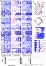
more conserved than surface exposed residues (Fig. 4a & Extended Data Fig. 7a,b).
The function landscape suggests the geometry of the designed cavity is critical to
activating DFHBI fluorescence: the key sequence features that specify the geometry
of the cavity - the glycine kinks and the tryptophan corner - are strictly conserved
(Fig. 4a). Among the seven coordinating residues from RIF docking, only a single substitution
(V103L) increased fluorescence (Fig. 4c, upper panel). Whereas the structure and function
landscapes were very similar at the bottom of the barrel (Fig. 4b), there was a striking
trade-off between stability and function at the top of the barrel around the designed
binding site (Fig. 4c): many substitutions that stabilize the protein drastically
reduce fluorescence activation (Fig. 4c, right). This bottom/top contrast indicates
that success in de novo design of fold and function requires a substantial portion
of the protein (in our case, the bottom of the barrel) to provide the driving force
for folding as the functional site will likely be destabilizing.
Guided by the comprehensive protein stability and fluorescence activation maps, we
combined substitutions at three positions that improved function without compromising
stability (V103L, V95AG and V83ILM; Extended Data Fig. 8a,b), and obtained variants
with 10-fold higher DFHBI fluorescence that form stable monomers without a disulfide
bond (b11L5F.1; Extended Data Fig. 8c). The crystal structure of one of the improved
variants (b11L5F_LGL; mutant 83L/95G/103L in Extended Data Fig. 8b) was solved to
2.2Å and was very close to the design model with the majority of the buried side chains
adopting the designed conformation (Extended Data Fig. 9a–d). However, the electron
density in the binding site could not be resolved, consistent with the multiple DFHBI
binding modes suggested by the docking calculations (Extended Data Fig. 9e–g; docking
calculations in Extended Data Fig. 5f). A second round of computational design calculations
was carried out to favor a specific binding mode by optimizing the protein-ligand
interactions in the lowest energy docked conformation, and to rearrange the hydrophobic
packing interactions in the bottom of the barrel now freed from the disulfide bond.
Five designs predicted by ligand docking calculations to have a single ligand binding
conformation were experimentally tested and three showed increased fluorescence activity,
the best of which increased the fluorescence by approximately 1.4-fold (b11L5F.2;
Extended Data Fig. 8d–e). Screening of two combinatorial libraries (based on b11L5F.1
and b11L5F.2) incorporating additional beneficial substitutions identified in the
b11L5F stability and function maps yielded variants with another 1.5-to-2 fold increased
fluorescence and improved protein stability (Extended Data Fig. 8f–h & 10a,b). We
refer to these mini- fluorescence-activating proteins as mFAPs in the remainder of
the text; mFAP0 and mFAP1 are variants of b11L5F.2, and mFAP2 of b11L5F.1. mFAP1 and
mFAP2 activate 0.5 μM DFHBI fluorescence by 80- and 60- fold with KD values of 0.56
μM and 0.18 μM, respectively (Fig. 5d).
The 1.8Å and 2.3Å crystal structures of mFAP0 and mFPA1 in complex with DFHBI were
virtually identical to the design models with an overall backbone RMSD of 0.91Å and
0.64Å (Fig. 5a–c & Extended Data Fig. 9h,i). DFHBI is in the planar Z conformation
with unambiguous electron density in both structures (Fig. 5a & Extended Data Fig.
9j). In addition to three designed hydrogen bonds, a water molecule was found to interact
with the solvent exposed phenol group in DFHBI (Fig. 5b). The DFHBI binding modes
in the crystal structures are nearly identical to the lowest-energy docked conformations
used in the second round of design calculations, with all-atom RMSD of 0.12Å and 0.35Å
respectively (Fig. 5c & Extended Data Fig. 9k). Three mutations shared by mFAP0 and
mFAP1 in the bottom barrel (P62D, M65L and L86MorY, Extended Data Fig. 10b) likely
stabilize the protein by helical capping and subtle hydrophobic rearrangements (Extended
Data Fig. 9l). The M27W mutation in mFAP1 introduced an additional hydrogen bond to
DFHBI that likely produces the 5nm red-shift in its fluorescence spectra (Fig. 5d;
Extended Data Fig. 10c,e). mFAP2, based on b11L5F.1, has a 6-residue insertion in
the seventh β-turn predicted to form multiple intra-loop hydrogen bonds (Extended
Data Fig. 10b, right).
In vivo fluorescence activation
To determine whether the designed DFHBI-binding fluorescence-activating proteins function
in living cells, we imaged mFAP1- and mFAP2-DFHBI complexes in E.coli, yeast, and
mammalian cells by conventional wide field epifluorescence microscopy and confocal
microscopy. Both mFAP1 and mFAP2 activated fluorescence in less than 5 minutes following
addition of 20µM DFHBI. Cytosolic expression of mFAPs in E.coli and mammalian cells
resulted in clear fluorescence throughout the cells (Fig. 5e & Extended Data Fig.
10f). Yeast cells with mFAPs targeted to the cell surface displayed fluorescence in
a thin region outside of the plasma membrane (Fig. 5f & Extended Data Fig. 10g). Fusion
of the mFAPs to a mitochondria-targeting signal peptide and to the ER localized protein
sec61β resulted in fluorescence tightly localized to these organelles in both fixed
(Fig. 5g&h) and living cells (Supplementary Videos) with a distribution comparable
to that of sec61β-GFP. The quantum yields of mFAP1 and mFAP2 in complex with DFHBI
are 2.0% and 2.1%, respectively (Extended Data Fig. 4g, comparable with Y-FAST:HBR
31
). The brightness of de novo mFAPs in complex with DFHBI is about 35-fold lower than
that of eGFP; there is still considerable room for improving their fluorescence activity.
Conclusion
It is instructive to compare the structures of our designed fluorescence-activating
proteins with those of natural fluorescent proteins (Fig. 6). Both are β-barrels,
and have similar chromophores, but our designs have less than half the residues and
narrower barrels connected with short β-turns (Fig. 6a). In both cases, specific protein-chromophore
interactions reduce energy dissipation from intramolecular motions
32
, but the hydrogen bonding and hydrophobic packing around DFHBI is different from
GFP and is tailored to the smaller and simpler β-barrel (Fig. 6b). The precise structural
control enabled by computational design, together with the greater exposure of the
chromophore, may prove useful for fluorescence-based imaging and sensing applications.
The comparison in figure 6 highlights the two primary advances in this paper: the
first successful de novo design of a β-barrel, and the first full de novo design of
a small molecule binding protein. The first advance required the elucidation of general
principles for designing β-barrels, notably the requirement for systematic symmetry
breaking to enable hydrogen bonding throughout the barrel structure. These principles,
identified by pure geometric considerations coupled with computer simulations following
failure of the initial parametric design approach, are borne out by both the crystal
structures and the sequence fitness landscapes. The second advance goes considerably
beyond the design of ligand binding proteins and catalysts to date, which has relied
on repurposing naturally occurring scaffolds. The three step approach described in
this paper — first, identifying the basic principles required for specifying a general
fold class, second, using these principles to generate a family of backbones with
pocket geometries matched to the ligand or substrate of interest, and third, designing
complementary binding pockets buttressed by a deeper hydrophobic core — provides a
general solution to the problem of de novo design of ligand-binding proteins. This
generative approach allows the exploration of an effectively unlimited set of backbone
structures with shapes customized to the ligand or substrate of interest and, equally
importantly, provides a critical test of our understanding of the determinants of
folding and binding that goes well beyond descriptive analyses of existing protein
structures.
Methods
Code availability.
The Rosetta macromolecular modelling suite (http://www.rosettacommons.org) is freely
available to academic and non-commercial users. Commercial licenses for the suite
are available via the University of Washington Technology Transfer Office. Design
protocols and analysis scripts used in this paper are available in the Supplementary
Information and on https://dx.doi.org/10.5281/zenodo.1216229. The source code for
RIF docking implementation is freely available at https://github.com/rifdock/rifdock.
Data availability.
The atomic coordinates and experimental data of BB1, b10, b11L5F_LGL, mFAP0-DFHBI,
and mFAP1-DFHBI crystal structures have been deposited in the RCSB Protein Database
with the accession numbers of 6D0T, 6CZJ, 6CZG, 6CZH, and 6CZI respectively. All the
design models, Illumina sequencing data, sequencing analysis and source data (Fig.2
&.4, Extended Data Fig. 6e, 7, 8a&h) are available on https://dx.doi.org/10.5281/zenodo.1216229.
Computational design of nonfunctional β-barrels.
De novo design of nonfunctional β-barrels can be divided into two main steps: backbone
construction and sequence design. For backbone construction, two different approaches
were presented: parametric backbone generation and fragment-based backbone assembly.
Example scripts and command lines for each method are available in Supplementary Data.
Parametric backbone generation and sequence design based on hyperboloid models.
β-strand arrangements were generated using the equation of a hyperboloid of revolution
with an elliptic cross-section, sampling the elliptic radii around the ideal value
of β-barrel radius with number of strands ‘n’ and the shear number ‘S’ (see Supplementary
Methods). Eight β-strands were arranged as equally spaced straight lines running along
the surface of the hyperboloid. A reference Cα was defined as the intersection between
the first strand and the cross-section ellipse. The other Cα were systematically populated
along the 8 strands from this reference residue. The peptide backbone was generated
from the Cα coordinates using the BBQ software
38
. The arrangements of discrete β-strands were minimized with geometric constraints
to favor backbone hydrogen bonds. One round of fixed-backbone sequence design calculation
was carried out to pack the barrel cavity with hydrophobic residues. The resulting
β-strand arrangements with the best hydrogen bond connectivity and the tightest hydrophobic
packing were selected to be connected by short (2 to 4 residues) β-turns. Two iterations
of the loop hashing protocol implemented in RosettaRemodel
39
were performed to close the strands and refine the turns. The sequence design of those
β-turns was constrained to sequence profiles derived from natural proteins. Low energy
amino acid sequences were obtained for the connected backbones using a flexible-backbone
design protocol (see Supplementary Data). Designs with high sequence propensity for
forming β-strands, reasonable peptide bond geometry, and tight-packed hydrophobic
cores are selected for experimental test (see Supplementary Table 2).
Backbone assembly from fragments guided by a 2D map.
The presented 2D map (Fig. 1d) was designed with the longest strand length observed
in soluble β-barrel structures to obtain a β-barrel tall enough for accommodating
a hydrophobic core and a binding cavity. The length of each strand depends on its
specific position and the shear number of the barrel (see Supplementary Methods).
Glycine kinks and β-bulges were placed on the map as described in the main text. Specific
β-turn types were used to connect the β-strands based on their relative positions
to β-bulges (see Supplementary Methods). Based on this 2D map, we generated a constraint
file and a blueprint file to guide the assembly of the barrel using peptide fragments
from Rosetta fragments library. In the constraint file, each backbone hydrogen bond
was described as a set of distance and angle constraints (Extended Data Fig. 5b).
A set of distance and torsion constraints specific to the tryptophan corner were added
to the constraint file (Extended Data Fig.3g–j, and Supplementary Methods). In the
blueprint file, a torsion angle bin was attributed to every residue in the peptide
chain, according to Rosetta’s ABEGO nomenclature. After minimizing the assembled backbones
using Rosetta centroid scoring function with imposed constraints, our protocol output
an ensemble of poly-valine β-barrel backbones with defined glycine kinks, β-bulges,
β-turns and the backbone of the tryptophan corner. The main challenge in building
scaffolds with this protocol is to achieve a good balance between the constraints
weight, structure diversity and backbone torsion angles. For this work, we circumvented
this problem by performing two additional rounds of sequence design calculation to
regularize and prepare scaffolds for designing ligand binding β-barrels (Extended
Data Fig. 5b–d and Supplementary Methods).
Sequence design of nonfunctional β-barrels.
500 poly-valine backbones with good hydrogen bonds and torsion angles were selected
as input for Rosetta sequence design. Low energy sequences for the desired β-barrel
fold were optimized over several rounds of flexible-backbone sequence design. We employed
a genetic algorithm to effectively search the sequence space: each parent backbone
was used as input to produce 10 designs through individual Monte Carlo searching trajectory.
The best ~10% of the output designs were selected based on the evaluation for total
energy, backbone hydrogen bonds, backbone omega and phi/psi torsion angles and hydrophobic
packing interactions. The selected models were used as inputs for the next round of
design calculation. After 12 rounds of design and selection, no more improvements
on the backbone quality metrics were observed (an indication of searching convergence).
We then performed a backbone refinement by minimization in Cartesian space and a final
round of design calculation (backbone flexibility was limited in torsion space for
all the design calculation). The final top designs converged to the offspring of 3
initial backbones, sharing 36% to 99% sequence identity. For every parent backbone,
one or two designs with the best hydrophobic packing interactions were selected for
experimental characterization. The four designs (BB1–4) share 46% to 72% sequence
identity.
Computational design of DFHBI-binding fluorescence-activating β-barrels.
DFHBI is short for chemical name ((Z)-4-(3,5-difluoro-4-hydroxybenzylidene)-1,2-dimethyl-1H-imidazol-5(4H)-one).
De novo design of DFHBI-binding β-barrels consists of three steps: 1) generation of
ensembles β-barrel scaffolds (see above), 2) ligand placement by RIF docking and 3)
sequence design. 200 input scaffolds were generated in step 1 and used in the following
steps. Example scripts and command lines are available in Supplementary Data.
Rotamer Interaction Field (RIF) docking.
The Rotamer Interaction Field (RIF) docking method performs a simultaneous, high-resolution
search of continuous rigid-body docking space as well as a discrete sequence design
space. The search is highly optimized for speed and in many cases, including the application
presented here, is exhaustive for given scaffold/ligand pair and design criteria.
RIF docking comprises two steps. In the first step, ensembles of interacting discrete
side chains (referred to as “rotamers”) tailored to the target are generated. Polar
rotamers are placed based on hydrogen bond geometry while apolar rotamers are generated
via a docking process and filtered by an energy threshold. All the RIF rotamers are
stored in ~0.5Å sparse binning of the 6-dimensional rigid body space of their backbones,
allowing extremely rapid lookup of rotamers that align with a given scaffold position.
To facilitate the following docking step, RIF rotamers are further binned at 1.0Å,
2.0Å, 4.0Å, 8.0Å and 16.0Å resolutions. In the second step, a set of β-barrel scaffolds
is docked into the produced rotamer ensembles, using a hierarchical branch-and-bound
search strategy (see Extended Data Fig. 5a). Starting with the coarsest 16.0Å resolution,
an enumerative search of scaffold positions is performed: the designable scaffold
backbone positions are checked against the RIF to determine whether rotamers can be
placed with favorable interacting scores. All acceptable scaffold positions (up to
a configurable limit, typically 10 million) are ranked and promoted to the next search
stage. Each promoted scaffold is split into 26 child positions in the 6D rigid body
space, providing a finer sampling. The search is iterated at 8.0Å, 4.0Å, 2.0Å, 1.0Å
and 0.5Å resolutions. A final Monte Carlo-based rotamer packing step is performed
on the best 10% of rotamer placements to find compatible combinations.
Sequence design of DFHBI-binding β-barrels.
A total number of 2,102 DFHBI-scaffold pairs from RIF docking were continued for Rosetta
sequence design. Our design protocol iterated between a fixed-backbone binding site
design calculation and a flexible-backbone design for the rest of scaffold positions.
Three variations of this design protocol were used during the sequence optimization.
In the initial two rounds of design calculation, RIF rotamers (interacting residues
placed during RIF docking) were fixed to maintain the desired ligand coordination.
Repacking of RIF rotamers was allowed in the final round of design calculation, assuming
that the binding sites have been optimized enough to retain these interactions. A
Rosetta mover that biases aromatic residues for efficient hydrophobic packing were
added after the first round of design. A similar selection approach and Cartesian
minimization as described for nonfunctional sequence design were used to propagate
sequence search and refine the design models. Evaluations on ligand binding interface
energy and shape complementarity were added to the selection criteria. The final set
of designs were naturally separated into clusters based on their original RIF docking
solutions. For each cluster, a sequence profile was generated to guide an additional
two rounds of profile-guided sequence design. 42 designs from 22 RIF docking solutions
(20 input scaffolds) were selected for experimental characterization (see Supplementary
Table 3).
Post-design model validation and ligand docking simulation.
To validate the protein and ligand conformations of the selected designs, we applied
model refinement followed by ligand docking simulation. Protein model refinement was
carried out on the unbound model of the designs by running five independent 10-ns
MD simulations followed by structural averaging and geometric regularization
40
. Then ligand docking simulation was performed on this refined unbound structure using
RosettaLigand
41
using Rosetta energy function
42
, allowing rigid body orientation and intra-molecular conformation of the ligand as
well as surrounding protein residues (both on side chains and backbones) to be sampled.
The ligand-binding energy landscapes were generated by repeating 2,000 independent
docking simulations.
Design of disulfide bonds.
The disulfide bonds were designed between the N-terminal 3–10 helix and a residue
on one of the β-strands on the opposite side to the tryptophan corner. The first 6
residues of the designs model were rebuilt with RosettaRemodel
39
and checked for disulfide bond formation using geometric criteria. Once a disulfide
bond was successfully placed, the N-terminal helix was redesigned.
Redesign of β-turns for b11.
Three β-turns (loop 3, 5 and 7) surrounding the DFHBI-binding site of b11 were redesigned
to make additional protein-ligand contacts. A set of “pre-organized” loops with high
content of intra-loop hydrogen bonds and low B-factors were collected from natural
β-barrel structures, and used as search template to build individual loop fragment
library. Those custom libraries were used as input for RosettaRemodel to build an
ensemble of loop insertions in the b11 design model bound to DFHBI. Two rounds of
flexible-backbone design calculation were carried out to optimize ligand interface
energy and shape complementarity using sequence profiles to maintain the template
backbone hydrogen bonds. Designed loop sequences were validated in silico by kinematic
loop closure
43
(KIC). 500 loop conformations were generated by independent KIC sampling and scored
by Rosetta energy function. 36 designs with improved ligand interface energy, shape
complementarity and converged loop sampling were selected for experimental characterization
(see Supplementary Data and Supplementary Table 6).
Redesign of β-barrel core and DFHBI-binding site for b11L5F.1.
After releasing the disulfide bond in b11L5F, with ligand modeled in the lowest-energy
docked conformation for b11L5F (see Extended Data Fig. 5f, right), we performed another
round of design calculation to further optimize the β-barrel core packing and ligand
binding interactions. The design protocol was very similar to the one used before
with fixed ligand hydrogen-bonding residues from RIF docking. 5 designs with 9–15
mutations after manual inspection were selected for experimental characterization.
Protein expression and purification.
Genes encoding the nonfunctional β-barrel designs (41 from parametric design and 4
from fragment-base design) were synthesized and cloned into the pET-29 vector (GenScript,
Inc). Plasmids were then transformed into BL21*(DE3) E. coli strain (NEB, Inc). Protein
expression was induced either by 1mM isopropyl β-d-thiogalactopyranoside (IPTG) at
18°C, or by overnight 37°C growth in Studier autoinduction medium. Cells were lysed
either by sonication (for 0.5–1L cultures) or FastPrep (MPBio, Inc) (for 5–50mL cultures).
Soluble designs were purified by Ni-NTA affinity resin (Qiagen, Inc) and monomeric
species were further separated by Akta Pure fast protein liquid chromatography (FPLC)(GE
Healthcare, Inc) using a Superdex 75 increase 10/300 GL column (GE Healthcare, Inc).
56 genes encoding DFHBI-binding designs were synthesized and cloned into pET-28b vector
(Gen9, Inc). Protein expression and purification were carried out in the same way.
Circular dichroism (CD).
Purified protein samples were prepared at 0.5mg/ml in 20mM Tris buffer (150mM NaCl,
pH 8.0) or PBS buffer (25mM phosphate, 150mM NaCl, pH7.4). Wavelength scans from 195nm
to 260nm were recorded at 25℃, 75℃, 95℃ and cooling back to 25℃. Thermal denaturation
was monitored at 220nm or 226nm from 25℃ to 95℃. Near-UV wavelength scan from 240nm
to 320nm and tryptophan fluorescence emission were recorded in the absence and presence
of 5M guanidinium chloride (GuHCl). Chemical denaturation in GuHCl was monitored by
both tryptophan fluorescence and near-UV CD signal at 285nm. The concentration of
the GuHCl stock solution was measured with a refractometer (Spectronic Instruments,
Inc). Far-UV CD experiments were performed on an AVIV model 420 CD spectrometer (Aviv
Biomedical, Inc). Near-UV CD and tryptophan fluorescence experiments were performed
on a Jasco J-1500 CD spectrometer (Jasco, Inc). Protein concentrations were determined
by 280nm absorbance with a NanoDrop spectrophotometer (ThermoScientific, Inc). Melting
temperatures were estimated by smoothing the sparse data with a Savitsky-Golay filter
of order 3 and approximating the smoothed data with a cubic spline to compute derivatives.
Reported Tm values are the inflection points of the melting curves.
Size Exclusion Chromatography with Multi-Angle Light Scattering (SEC-MALS).
Protein samples were prepared at 1–3mg/ml and applied to a Superdex 75 10/300 GL column
(GE Healthcare) on a LC 1200 Series HPLC machine (Agilent Technologies, Inc) for size-based
separation, followed by a miniDAWN TREOS detector (Wyatt Technologies, Inc) for light-scattering
signals.
Fluorescence binding assay.
Protein-activated DFHBI fluorescence signals were measured in 96-well plate format
(Corning 3650) on a Synergy neo2 plate reader (BioTek, Inc) with λex = 450nm or 460nm
and λem = 500nm or 510nm. Binding reactions were performed at 200µL total volume in
PBS pH7.4 buffer. Protein concentrations were determined by 280nm absorbance as described
above. DFHBI (Lucerna, Inc) were resuspended in DMSO as instructed to make 100mM stock
and diluted in PBS to 0.5–10µM.
Library construction.
Deep mutational scanning library for b11L5F were constructed by site-directed mutagenesis
as described
44
. 111 PCR reactions were carried out using DNA oligos directed to each position in
two 96-well polypropylene plates (USA Scientific, 1402–9700), and products were pooled
and purified by gel extraction kit (Qiagen, Inc) for yeast transformation. Combinatorial
libraries for b11L5F.1 and b11L5F.2 were assembled using synthesized DNA oligos (Integrated
DNA technologies, Inc) as described
45
. Selected positions were synthesized with 1–2% mixed bases to control mutation rate
and library size. Full-length assembled genes were amplified and purified for yeast
transformation as described
46
.
Yeast surface display and fluorescence activated cell sorting (FACS).
Transformed yeast cells (strain EBY100)
46
were washed and re-suspended in PBSF (PBS plus 1g/L of BSA). DFHBI in DMSO stock was
diluted in PBSF for labeling yeast cells at various concentrations. PBSF-treated cells
were incubated with DFHBI for 30 min to 1 hour at room temperature on a benchtop rotator
(Fisher Scientific, Inc). Library selections were conducted using GFP fluorescence
channel at 520nm with 488nm laser on a SH800 cell sorter (Sony, Inc). Proteolysis
treatment and fluorescence labelling were performed in the same way as described
30
. Cell sorting parameters and statistics for all selections are given in Supplementary
Table 16.
Deep sequencing and data analysis.
Pooled DNA samples for b11L5F deep mutational scanning library were transformed twice
to obtain biological replicates. Two libraries were treated and sorted in a parallel
fashion. Yeast cells of naive and selected libraries were lysed and plasmid DNA was
extracted as described
47
. Illumina adaptor sequences and unique library barcodes were appended to each library
by PCR amplification using population-specific primers (see Supplementary Table 8).
DNA was sequenced in paired-end mode on a MiSeq Sequencer (Illumina, Inc) using a
300-cycle reagent kit (Catalog number: MS-102–3003). Raw reads were first processed
using the PEAR program
48
and initial counts analysed with scripts adapted from Enrich
49
. Stability scores were modeled using sequencing counts from proteolysis sorts as
described
30
. Unfolded states were modeled without disulfide bonds (Cysteine were replaced by
Serine). Function scores were modeled using sequencing counts from DFHBI fluorescence
sorts. A simple meta-analysis statistical model with a single random effect was applied
to combine two replicates using the framework developed in Enrich2
50
.
BB1 crystal structure.
BB1 protein was concentrated to 20 mg/ml in an AMICON Ultra-15 centrifugation device
(Millipore, Inc), and sequentially exchanged into 20mM Tris pH8.0 buffer. The initial
screening for crystallization conditions was carried out in 96-well hanging drop using
commercial kits (Hampton Research, Inc & Qiagen, Inc) and a mosquito (TTP LabTech).
With additional optimization, BB1 protein crystallized in 0.1 M BIS-Tris pH 5.0 and
2M ammonium sulfate at 25℃ by hanging drop vapor diffusion with 2:1 (protein: solution)
ratio. Diffraction data for BB1 was collected over 200° with 1° oscillations, 5s exposures,
at the Advanced Light Source (Berkeley, CA) beamline 5.0.1 on an ADSC Q315R area detector,
at a crystal-to-detector distance of 180mm. The data was processed in space group
P21 to 1.63 Å using Xia2
51
. The BB1 design model was used as a search model for molecular replacement using
the program Phaser
52
, which produced a weak solution (TFZ 6.5). From this, a nearly complete model was
built using the Autobuild module in Phenix
53
. This required the rebuild-in-place function of autobuild to be set to “False”. Iterative
rounds of model building in the graphics program Coot
54
and refinement using Phenix.refine
55
produced a model covering the complete BB1 sequence. Diffraction data and refinement
statistics are given in Supplementary Table18.
b10, b11L5F_LGL crystal structure and mFAPs-DFHBI co-crystal structures.
b10 was initially tested for crystallization via sparse matrix screens in 96-well
sitting drops using a mosquito (TTP LabTech). Crystallization conditions were then
optimized in larger 24-well hanging drops. b10 crystallized in 100mM HEPES pH 7.5
and 2.1M Ammonium sulfate at a concentration of 38 mg/mL. The crystal was transferred
to a solution containing 0.1 M HEPES pH 7.5 with 3.4 M Ammonium sulfate and flash
frozen in liquid nitrogen. Data was collected with a home-source rotating anode on
a Saturn 944+ CCD and processed in HKL2000
56
.
b11L5F_LGL was concentrated to 19.6 mg/mL (1.58 mM), incubated at room temperature
for 30 minuets with 1 mM TCEP then mixed with an excess of DFHBI (re-suspended in
100% DMSO). b11L5F_M11 complexed with DFHBI was screened via sparse matrix screens
in 96-well sitting drops using a mosquito (TTP LabTech) and crystallized in 100 mM
Bis-Tris pH6.5 and 45% (v/v) Polypropylene Glycol P 400. The crystal was flash frozen
in liquid nitrogen directly from the crystallization drop. Data was collected with
a home-source rotating anode on a Saturn 944+ CCD and processed in HKL2000
56
.
mFAP0 and mFAP1 were mixed with excess DFHBI (re-suspended in 100% DMSO), while keeping
the final DMSO concentration at less than 1%. The mFAP0 and mFAP1 complexes were then
concentrated to approximately 41 mg/mL and 64 mg/mL, respectively, and initially tested
for crystallization via sparse matrix screens in 96-well sitting drops using a mosquito
(TTP LabTech). Crystallization conditions were then optimized in larger 24-well hanging
drops macroseeded with poor quality crystals obtained in sitting drops. mFAP0 complexed
with DFHBI crystallized in 200 mM Sodium chloride, 100 mM HEPES pH 7.5 and 25% (w/v)
Polyethylene Glycol 3350. The crystal was transferred to the mother liquor plus 2
mM DFHBI and 10% (w/v) Polyethylene Glycol 400 then flash frozen in liquid nitrogen.
Data was collected at the Berkeley Center for Structural Biology at the Advanced Light
Source (Berkeley, CA), on beamline 5.0.2 at a wavelength of 1.0 Å. and processed in
HKL2000
56
. mFAP1 complexed with DFHBI crystallized in 100 mM MES pH6.5 and 12% (w/v) Polyethylene
Glycol 20,000. The crystal was transferred to the mother liquor plus 2 M DFHBI and
15% glycerol then flash frozen in liquid nitrogen. Data was collected with a home-source
rotating anode on a Saturn 944+ CCD and processed in HKL2000
56
.
Structures were solved by Molecular Replacement with Phaser
52
via phenix
53
using the Rosetta design model with appropriate residues cut back to C-alpha and DFHBI
removed. The structure was then built and refined using Coot
54
and phenix
55
, respectively, until finished. Diffraction data and refinement statistics are given
in Supplementary Table18.
Statistics and reproducibility
(Fig. 1c) The models were colored based on the mean values of repulsion energy by
position (Rosetta fa_rep) derived from a set of polyvaline backbones relaxed with
constraints (n=189 independently generated models); relaxed with constraints with
a glycine in the middle of each Cβ-strip (n=186 independently generated models) and
relaxed without constraints with glycines and β-bulges (n=194 independently generated
models). This experiment has been performed twice on different sets of backbones and
produced similar results. (Fig. 2b–e) BB1 was purified and sized with SEC at least
5 times independently, yielding different ratio of monomeric to oligomeric species
(20%−75%). The fraction of monomer could be increased by heat-shocking the cells at
42°C shortly before induction. Two biological replicates of the far and near UV CD
and tryptophan fluorescence spectra acquisition of BB1 were done with similar results,
and the chemical denaturation experiment performed once. (Extended Data Fig.4a–c)
The analysis of BB1 with SEC-MALS was repeated twice on independently prepared protein
samples and similar molecular weights were obtained. Additionally, the experiments
were repeated on one sample stored at 4°C at different time points (t=0; t=7 days
and t=30 days); all experiments had similar results and confirmed the stability of
the monomeric species. BB2,3 and 4 were purified once. The molecular weight (with
SEC-MALS) and the far UV CD spectra of the purified proteins were tested one time.
The sizing of purified BB1 mutants was performed once, with WT BB1 as an internal
control.
Cell Culture and Transfection
COS-7 cells (ATCC CRL-1651) were grown in DMEM supplemented with 1x NEAA, 100 units/mL
penicillin, 100 µg/mL streptomycin, and 10% FBS; and harvested using 0.25% Trypsin
EDTA. Per transfection, approximately 1 million cells were transfected with 2 µg of
plasmid using 18 µL of Lonza SE cell supplement, 82 µL of Lonza SE nucleofection solution
and pulse code DS-120 on a Lonza 4D X Nucleofector system. After nucleofection cells
were immediately seeded into ibidi µ-Slide 8 well glass bottom chambers at a density
of ~30,000 cells/well and incubated overnight at 37 °C.
Cell Fixation
Cells were fixed at 37°C for 10 minutes in PFA/GA fixation solution containing 100
mM aqueous PIPES buffer pH 7.0, 1 mM EGTA, 1 mM MgCl2, 3.2% paraformaldehyde, 0.1%
glutaraldehyde; reduced for 10 minutes with freshly prepared 10 mM aqueous sodium
borohydride; then rinsed with PBS for 5 minutes.
Microscopy
Conventional widefield epifluorescence imaging was performed on an inverted Nikon
Ti-S microscope configured with a 60 × 1.2 NA water-immersion objective lens (Nikon,
Melville, NY, USA), a light emitting diode source (LED4D120, Thorlabs, Newton, NJ,
USA), a multiband filter set (LF405/488/532/635-A-000, Semrock, Rochester, NY, USA)
and images were captured with a Zyla 5.5 sCMOS camera (Andor, Windsor, CT, USA). The
samples were illuminated 470 nm light at an intensity of ~2 W/cm2 and with 200 ms
exposures. For live cell experiments, samples were incubated at 37°C with Gibco CO2
Independent Medium containing 50 µM DFHBI for 10 minutes prior to imaging. Time lapse
movies were acquired over a period of 5 minutes with a 200 ms exposure every 5 seconds.
For fixed cell imaging, samples were incubated at room temperature (~22°C) in PBS
containing 50 µM DFHBI for 10 minutes prior to imaging.
Extended Data
Extended Data Figure 1:
Parametric design: workflow and shortcomings.
a, Schematic representation of the parametric approach to generate β-barrel designs.
b-d, Comparison between β-barrels of type (n=8;S=8, b), type (n=8;S=10, c) and type
(n=8;S=12, d); showing an example of 2D map with residue connectivity (top), the arrangement
of the Cβs in the Cβ-strips (middle) and the packing pattern of the core side-chains
(bottom). The difference in shear number translates into different overall strand
staggering and barrel radii. The number of core Cβ-strips (top, middle) results in
different arrangements of side-chains in the core of the barrel. e&f, The parametric
designs exhibited distorted hydrogen bonds, reflected by the shear distance (defined
in e) between paired antiparallel β-strands residues. The shear distance in the design
deviate from the distribution observed in native β-sheet proteins (f).
Extended Data Figure 2:
Glycine kinks release strain in β-barrel backbones.
a, Fraction of retained hydrogen bond interactions after relaxation with Rosetta (‘relax’)
of uniform polyvaline backbones (white) and polyvaline backbones with a glycine in
the middle of each Cβ-strip (grey). We compare disconnected strand arrangements generated
with the parametric hyperboloid model (n=225 independently generated models), the
cylindric model (n=36 independently generated models), the coiled-coil model (n=150
independently generated models) and assembled based on a 2D map (n=144 independently
generated models). Center line, median; box limits, upper and lower quartiles; whiskers,
minimum and maximum values; points, outliers. b&c, In polyvaline backbones (n=189
independently generated models) relaxed with constraints to maintain hydrogen bonds
between strands, several residues have unfavourable left-handed twist (c). The local
strand twist is calculated on a sliding window of 4 residues along β-strands, as the
angle between the vectors Cα1- Cα3 and Cα2- Cα4. The handedness of the twist is defined
as the triple scalar product between these two vectors and the central axis of the
barrel. Positive and negative values denote right-handed and left-handed twist, respectively.
(b). d, After relaxation (‘FastRelax’), the valine positions in the middle of each
Cβ-strip remained in the β-sheet specific ABEGO space (right); or were shifted towards
the positive Φ space (E ABEGO) if mutated to glycines (bottom). e, A similar torsion
angle distribution was observed for glycines in the β-strands of native β-barrels
(n=35 high resolution crystal structures). f, In comparison with regular β-strands
(top), the presence of glycine kinks (bottom) increases the local bending of the strands
and creates corners in an otherwise circular barrel cross-section. g, The bending
angle α is calculated on a sliding window of 3 residues.
Extended Data Fig. 3:
Placement of β-bulges, β-turns and the tryptophan corner.
a, Change of curvature (from convex to concave) and protrusion (dashed circle) of
the longest hairpin associated with the placement of a glycine kink at position 44.
b, relationship between the “corners” in the β-sheet (dashed line) generated by the
glycine kinks and the type and position of the β-bulges and β-turns (Supplementary
methods). Cα are shown as spheres and colored by ABEGO type. The bottom of the barrel
was defined as the side of the N- and C-termini. c, The type I β-turn (‘AA’ ABEGO
type) is frequently found at the second position relative to a β-bulge in native proteins
and was selected to connect bottom hairpins. d, This choice is further supported by
the enrichment of type I (AA) turns over the canonical type I’ turn (GG) in native
β-barrels (n=35 high resolution crystal structures). e&f, Poly-valine backbones built
with β-bulges and the corresponding β-turns (n=194 independently generated models)
retain more hydrogen bonds after relaxation than backbones built without β-bulges
and with canonical type I’ β-turns (n=186 independently generated models) (e) and
exhibit better scored hydrogen bonds per β-strand residue flanking the β-turns (f).
Center line, median; box limits, upper and lower quartiles; whiskers, minimum and
maximum values; points, outliers. g, Superposition of tryptophan corner motifs (n=41
high resolution crystal structures) extracted from native β-barrels. h-j, Amino acid
preference and torsional constraints derived from the set and used to model the tryptophan
corner. Bounded constraints limits are shown as dashed lines.
Extended Data Fig. 4:
Biochemical and structural characterizations of designs BB1–4.
a, Results of experimental characterization of the nonfunctional designs (BB1–4).
Reproducibility is described in the Methods. †E-value is calculated by BLAST the non-redundant
protein database. b, Far-UV CD spectra of designs BB2 and BB3 at 25°C. c, SEC-MALS
analysis showed a major monomer peak for BB1 and a major tetramer peak for BB2. d,
Variants of BB1 with residues of the tryptophan corner and glycine kinks mutated to
alanine were purified and sized. SEC are superimposed to the SEC trace of wild-type
BB1 (WT). The mutations of all residues of the tryptophan corner eliminate the monomeric
peak. Most of the glycine kink mutations negatively affect the monomeric species.
The exceptions are Gly53 and Gly55, which are following each other on the fourth strand.
Only one glycine kink per strand might be sufficient to introduce enough negative
twist to un-strain the β-barrel. e-f, Deviations between BB1 design model and crystal
structure. (e) One of the three bottom turns of the crystal structure (grey) significantly
deviates from the design model (magenta) and forms additional crystal contacts (indicated
by a dashed circle). (f) Three phenylalanine side-chains have different rotameric
states. In the crystal structure, Phe41 interacts with Gly53 (which shows the most
backbone deviation between the crystal structure and the design) to form an aromatic
rescue motif
34
. It is likely that the Phe rotamers discrepancy reflect a scoring/sampling challenge
to accurately capture such aromatic rescue; MD simulation starting from the crystal
structure (cyan) was also unable to recover the correct Phe41-Gly53 interaction. g,
Biophysical properties (absorbance/fluorescence spectra, quantum yield and binding
affinity) of mFAP1 and mFAP2 in complex with DFHBI. Average values from three biological
replicates were used for the nonlinear regression to determine the KD. The error estimates
are the standard deviation from the fitting calculation. *λabs is peak absorbance
wavelength, λex is peak excitation wavelength and λem is peak emission wavelength.
†Absolute quantum yield is measured with an integrating sphere; Relative quantum yield
is measured using acridine yellow and fluorescein as the standards. ‡reported value
26
. §From
37
. ||From
31
.
Extended Data Fig. 5:
RIF docking grid-based search algorithm, β-barrel scaffold construction and post-design
ligand docking simulations.
a, Illustration of grid-based hierarchical search strategy in RIF docking. After generating
an ensemble of interactions for the target ligand (Figure 3), each one of the selected
scaffold is docked into the fixed “rotamer interaction field (RIF)” using the grid-based
hierarchical searching algorithm. This search procedure starts from coarse sampling
grids to fine sampling grids in 3D space. An example 2D grid scheme is shown in the
upper row, from the lowest resolution (coarse sampling, left) to the highest resolution
(fine sampling, right). At each searching stage, the backbone is assigned to different
grids based on its relative position and the resulting docking configurations are
scored. The top-scored backbone positions (highlighted by cyan circles in the 2D scheme)
are shown as 3D structures in the lower row for each searching resolution and are
continued for the next grid search and scoring. The 3D structure example shown here
was streptavidin structure (PDB ID: 1STP) with grid searching resolutions of 8.0Å,
4.0Å, 2.0Å, and 1.0Å. b-d, β-barrel scaffold construction for small molecule binding.
Three geometric constraints (b) were used to describe each backbone hydrogen bond
and drive the backbone assembly during Rosetta low-resolution centroid modeling. Backbones
generated with all three constraints had a very narrow Φ/Ψ distribution as a result
of strong constraints (c, Ramachandran plot in upper left, Set 1, density colored
in blue); by omitting N-H-O angle constraint, backbone torsion diversity slightly
improved (c, upper right, Set 2). These two raw backbone sets yielded few non-redundant
RIF docking solutions (d, blue bars). After two rounds of sequence design calculation
using Rosetta full-atom force field (Supplementary Methods), regularized backbones
(peptide bonds with proper dihedral geometry) and broadened Φ/Ψ distribution (c, Ramachandran
plot in the lower row, density colored in orange) yielded more unique RIF docking
solutions (d, orange bars). e, Computed metrics for 42 designs ordered and tested.
Results from ab initio folding simulation were scaled to 0.0 to 1.0, with 1.0 represents
a funnel-shaped folding landscape
35
. f, Alternative ligand binding conformations revealed by post-design ligand docking
simulations. The lowest-energy docking conformation using the design model (by simply
taking out the ligand from the pocket) was similar to the designed DFHBI-binding mode
(top left, grey; designed binding mode was circled in grey in the energy landscape
in the lower row). Docking simulations using MD-refined the apo protein model revealed
an alternative equal-energy docking conformation (top right, green) indicated by a
green circle in the docking energy landscapes (lower row). Both binding modes rely
on three hydrogen bonding residues from RIF docking (upper row).
Extended Data Fig.6:
Biochemical and structural characterization of design b10, b32, b11.
a, Size-exclusion chromatogram (SEC) of His6-tagged b10 and b32 after Ni-NTA affinity
purification. The monodispersed peaks of absorbance at 280nm of b10 and b32 (cyan
and lavender, respectively) have an elution volume compatible with the monomeric β-barrel
(14kDa), based on their relative position to the protein standard peaks (dashed line).
n biological replicates were performed with similar observation: n=4 for b10, n=5
for b32. b, Comparison of ligand binding pocket in b10 design model (middle, grey)
with the crystal structure (left, cyan). The side-chain disagreements are highlighted
with a dashed black circle on the right panel. c&d, The designed disulfide bond as
a stabilizing mechanism. SEC curves of His6-tagged b11 (purple line) and b38 (dark
yellow line) were overlaid to show the appearance of a monomer peak for b11 (the same
standard as in a was applied here). A disulfide bond connecting the N-terminal helix
to a β-strand (Q1C and M59C, circled in d) along with four mutations of neighboring
residues, were introduced into design b38 (dark yellow) to make design b11 (purple).
n biological replicates were performed with similar observation: n=3 for b38, n=5
for b11. e, Far-UV circular dichroism(CD) spectra of b10, b32 and b11. Left: spectra
at different temperatures within one heating-cooling cycle; Right: thermal melting
curves (b10’s CD signal was monitored at 220nm; b32 and b11 at 226nm). b11 likely
forms an amyloid-like beta structure at 95°C (left, bottom row) with a negative peak
around 226nm
36
and refolds back after cooling to 25°C. The thermal stability of b11 decreases when
the disulfide was reduced with 1mM tris(2-carboxyethyl) phosphine (TCEP) (right, bottom
row). Measurements were performed once for each design (n=1). f, Fluorescence emission
spectra of b32, b11 and b11L5F in complex with DFHBI. With 200 µM proteins, b32, b11
and b11L5F can activate 10 µM DFHBI fluorescence by 8-, 12- and 18-fold, respectively.
n=2 biological replicates were performed with similar results. g, The residues designed
to interact with DFHBI contribute to b11 and b32 activity. Single or double knockouts
of hydrogen bonding residues (Y71, S23, N17 and T95) and a hydrophobic packing residue
(M15) showed decreased fluorescence intensity at 500nm in comparison with the wild-type
b11 or b32 (WT). Mutants were purified once for activity measurement. h&i, Re-designed
5-residue fifth turn in b11L5F. The original bulge-containing “AAG” β-turn in b11
(Extended Data Fig. 3b) was redesigned into a 5-residues turn. b11L5F was detected
by yeast surface display and flow cytometry (i and Supplementary Data). Yeast cells
displaying b11 and b11L5F showed an increased 520nm fluorescence signal (excited by
488nm laser, i). n=3 biological replicates were performed with similar observation.
Extended Data Fig. 7:
Deep mutational scanning maps for b11L5F.
a, The complete function (left) and protease stability (middle and right) landscapes
of b11L5F. Fluorescence activation scores, trypsin and chymotrypsin stability scores
were calculated as described in Supplementary Methods and demonstrated in the Supplemented
Data (b11L5F_DMS_analysis.ipy). n=2 biological replicates with >10-fold sequencing
coverage. Red color represents beneficial effect while mutations colored in blue color
are detrimental (relative to the wild-type b11L5F). Wild-type residues at each position
are indicated by black dots. b, b11L5F backbone model colored by the average stability
scores. Glycine backbone Cα are shown as spheres. c&d, Mutational scanning maps of
glycine kinks (G25, G43, G53, G55, G81 and G105) and tryptophan corner positions (G9,
W9 and R109) (c), and of glycines in the β-turns and prolines (d). e, Statistics of
the fluorescence activation and stability scores. The standard deviation between the
two replicates used for calculating fluorescence activation scores is smaller than
2 for most the data points (left); 95% confidence interval calculated for the proteolysis/stability
analysis is less than 0.25 for most the experimental protease EC50 values (middle
and right).
Extended Data Fig. 8:
Experimental and computational improvement based on b11L5F.
a-c, Incorporation of point mutations from deep mutational scanning. Beneficial mutations
that improve fluorescence activity without compromising protein stability (positive
scores relative to wild-type b11L5F; a, left, n=2 biological replicates) were mapped
onto b11L5F backbone model (a, right). Purified b11L5F variants incorporating those
single, double or triple mutations showed consistently improved fluorescence activity
(b). Binding titration curves were obtained for all six possible triple mutants (b,
right, n=1 biological measurement). b11L5F with V103L, V95A, V83I, C59V and C1S were
renamed as “b11L5F.1” (c). d, Characterization of five designs from the second round
of design calculation. Three of the five designs (nC1–5) based on b11L5F showed improved
binding activities by titrating purified proteins into 0.5µM DFHBI (d, n=1 biological
sample was used for the measurement). The best variant (nC5) was renamed as “b11L5F.2”.
e, Ligand docking simulations with the MD-refined apo b11L5F.2. Energy landscape was
plotted by comparing all the docking conformations to the design model (left). The
lowest-energy docking conformations (highlighted in green circle) match the design
model (right, design mode in silver and docking model in green). f&g, Characterization
of three best variants (mFAP0–2) from combinatorial library selections. Yeast cells
displaying mFAP proteins incubated with 5µM DFHBI analyzed by flow cytometry (f, excited
by 488nm laser, n=1 biological sample was used for the measurement with proper controls).
Purified proteins showed up to 100-fold fluorescence activation (5µM protein + 0.5µM
DFHBI, excited at 450nm and monitored at 500nm and 510nm in a plate reader, n=1 biological
measurement). h, Far-UV circular dichroism(CD) characterization of b11L5F.1, b11L5F.2,
mFAP0, mFAP1 and mFAP2. Left: spectra at different temperatures within one heating-cooling
cycle; Right: thermal melting curves (CD signals were monitored at 226nm, spectra
were recorded once (n=1) with internal noise estimation).
Extended Data Fig. 9:
Crystal structure of b11L5F_LGL, mFAP0 and mFAP1.
a-g, b11L5F_LGL crystal structure. (Protein samples of all six triple mutants in Extended
Data Fig. 8b(right) were prepared for crystallization. b11L5F_LGL with V83L/V95G/V103L
combination was successfully crystallized). Crystal contacts between protein copies
in one asymmetric unit (yellow) were mediated by two tyrosines (stick representation,
grey dashed circle); contacts between three asymmetric units (yellow, blue and green)
were formed between β-turns (black dashed circle), which might have displaced one
of the top β-turns (c). Overall backbone and side chain conformations in the design
model matched the crystal structure with a backbone Cα RMSD of 1.02Å (b, crystal in
yellow and design model in silver), and the designed disulfide bond was present in
the crystal structure (d). Ligand density in the crystal structure was ambiguous:
2Fo − Fc omit map showing the electron density after refinement without placing DFHBI
(e), the best ligand placement to match the density (f), and designed ligand binding
interactions (silver) overlaid with the crystallized binding pocket (g). h&i, Crystal
contacts in the DFHBI-bound structures of mFAP0(h) and mFAP1(i). Contacts between
proteins copies in one asymmetric unit were formed around 40V and 54Y (grey dashed
circle) that were introduced for helping crystallization (Extended Data Fig. 10a).
Contacts between asymmetric units were formed between β-turns (black dashed circle).
j, 2Fo−Fc omit electron density of DFHBI in the mFAP0-DFHBI complex crystal structure.
DFHBI density contoured at 1.0σ is clear and matches the planar conformation of the
ligand (right). k, Superposition of mFAP0 design model (silver) and the crystal structure
(magenta). Hydrogen bonds were indicated with dashed lines. e, Helical capping interactions
mediated by P62D mutation in mFAP1 crystal structure.
Extended Data Fig. 10:
Mapping of mutations introduced into b11 to yield the final brighter variants, biophysical
characterization of mFAP1&2, and epifluorescent images. a, Sequence alignment of b11-based
DFHBI-binding fluorescence-activating proteins.
Orange boxes indicate mutations or loop insertions introduced by computational design;
purple boxes highlight mutations rationally introduced based on the deep mutational
scanning maps (Extended Data Fig. 7&8); green boxes indicate mutations or loop insertions
that were incorporated during combinatorial library selections; K40V and K54Y in light
blue boxes were introduced to help crystal formation (Extended Data Fig. 9h&i). Despite
having hydrophobic residues on the surface, mFAP2 remains soluble at 150mg/mL. b,
mFAPs mutations mapped on the design models. Common mutations in all three mFAPs were
highlighted in bold. c, Absorbance spectra for DFHBI, mFAP1- and mFAP2-DFHBI complexes
(n=4 biological replicates with similar observation). d, Extinction coefficient determination
for DFHBI at 418nm. e, Normalized absorbance and fluorescence spectra of mFAP1- and
mFAP2-DFHBI complex (n=2 biological replicates with similar observation). f&g, Widefield
epifluorescence (bottom) and brightfield (top) images of E.coli and yeast cells with
20µM DFHBI. Untransformed E.coli Lemo21 cells (f, left, n=2 biological replicates
with similar observation) and yeast EBY100 cells displaying ZZ domain (g, left, n=2
biological replicates with similar observation) were treated with the same amount
of DFHBI and imaged in the same way (1000mA 470nm LED and 200ms exposure time).
Supplementary Material
1
2
3
Video 1
Video 2
Related collections
Most cited references49
- Record: found
- Abstract: found
- Article: not found
The coming of age of de novo protein design.
Po-Ssu Huang, Scott Boyken, David. Baker (2016)
- Record: found
- Abstract: found
- Article: not found
Simultaneous Optimization of Biomolecular Energy Functions on Features from Small Molecules and Macromolecules.
Hahnbeom Park, Philip Bradley, Per Greisen … (2016)
- Record: found
- Abstract: not found
- Article: not found
Global analysis of protein folding using massively parallel design, synthesis, and testing
Tamuka Chidyausiku, Inna Goreshnik, Alex T Ford … (2017)