13
views
0
recommends
+1 Recommend
0 collections
    0
    shares
      • Record: found
      • Abstract: found
      • Article: not found

      The role of HYAL2 in LSS-induced glycocalyx impairment and the PKA-mediated decrease in eNOS–Ser-633 phosphorylation and nitric oxide production

      research-article

      Read this article at

      ScienceOpenPublisherPMC
      Bookmark
          There is no author summary for this article yet. Authors can add summaries to their articles on ScienceOpen to make them more accessible to a non-specialist audience.

          Abstract

          This study examines the mechanism of glycocalyx injury under low shear stress (LSS). LSS-induced changes of eNOS phosphorylation and NO production are based on the deficiency of the glycocalyx barrier.

          Abstract

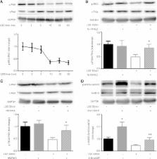
          Hyaluronan (HA) in the endothelial glycocalyx serves as a mechanotransducer for high-shear-stress–stimulated endothelial nitric oxide synthase (eNOS) phosphorylation and nitric oxide (NO) production. Low shear stress (LSS) has been shown to contribute to endothelial inflammation and atherosclerosis by impairing the barrier and mechanotransduction properties of the glycocalyx. Here we focus on the possible role of hyaluronidase 2 (HYAL2) in LSS-induced glycocalyx impairment and the resulting alterations in eNOS phosphorylation and NO production in human umbilical vein endothelial cells (HUVECs). We show that LSS strongly activates HYAL2 to degrade HA in the glycocalyx. The dephosphorylation of eNOS–Ser-633 under LSS was triggered after HA degradation by hyaluronidase and prevented by repairing the glycocalyx with high–molecular weight hyaluronan. Knocking down HYAL2 in HUVECs protected against HA degradation in the glycocalyx by inhibiting the expression and activity of HYAL2 and further blocked the dephosphorylation of eNOS–Ser-633 and the decrease in NO production in response to LSS. The LSS-induced dephosphorylation of PKA was completely abrogated in HYAL2 siRNA–transfected HUVECs. The LSS-induced dephosphorylation of eNOS–Ser-633 was also reversed by the PKA activator 8-Br-cAMP. We thus suggest that LSS inhibits eNOS–Ser-633 phosphorylation and, at least partially, NO production by activating HYAL2 to degrade HA in the glycocalyx.

          Related collections

          Most cited references31

          • Record: found
          • Abstract: found
          • Article: not found

          The endothelial glycocalyx: composition, functions, and visualization

          This review aims at presenting state-of-the-art knowledge on the composition and functions of the endothelial glycocalyx. The endothelial glycocalyx is a network of membrane-bound proteoglycans and glycoproteins, covering the endothelium luminally. Both endothelium- and plasma-derived soluble molecules integrate into this mesh. Over the past decade, insight has been gained into the role of the glycocalyx in vascular physiology and pathology, including mechanotransduction, hemostasis, signaling, and blood cell–vessel wall interactions. The contribution of the glycocalyx to diabetes, ischemia/reperfusion, and atherosclerosis is also reviewed. Experimental data from the micro- and macrocirculation alludes at a vasculoprotective role for the glycocalyx. Assessing this possible role of the endothelial glycocalyx requires reliable visualization of this delicate layer, which is a great challenge. An overview is given of the various ways in which the endothelial glycocalyx has been visualized up to now, including first data from two-photon microscopic imaging.
            Bookmark
            • Record: found
            • Abstract: found
            • Article: not found

            Endothelial cell glycocalyx modulates immobilization of leukocytes at the endothelial surface.

            A thick endothelial glycocalyx provides the endothelial surface with a nonadherent shield. Oxidized LDL (Ox-LDL) degrades the endothelial glycocalyx. We hypothesized that glycocalyx degradation stimulates leukocyte-endothelial cell adhesion, whereas intravascular supplementation with sulfated polysaccharides reconstitutes the endothelial glycocalyx and attenuates Ox-LDL-induced leukocyte-endothelial cell adhesion. Degradation of the endothelial glycocalyx by local microinjection of heparitinase (10 to 50 U/mL) into mouse cremaster venules dose-dependently increased the number of adherent leukocytes. Systemic administration of Ox-LDL (0.4 mg/100 g body weight) induced 10.1+/-0.9 adherent leukocytes/100 microm at 60 minutes. In the venules perfused with 500-kDa dextran sulfate (1 mg/mL), the number of adherent leukocytes at 60 minutes after Ox-LDL bolus application was not influenced (9.2+/-1.0 leukocytes/100 microm). However, the venules locally perfused with heparan sulfate (10 mg/mL) or heparin (1 mg/mL) displayed a significantly lower number of adherent leukocytes induced by Ox-LDL: 5.1+/-0.7 and 5.4+/-0.9 leukocytes/100 microm, respectively (P<0.05). Fluorescently labeled heparan sulfate and heparin, but not dextran sulfate, attached to the venule luminal surface after Ox-LDL administration. Endothelial glycocalyx degradation stimulates leukocyte immobilization at the endothelial surface. Circulating heparan sulfate and heparin attach to the venule wall and attenuate Ox-LDL-induced leukocyte immobilization.
              Bookmark
              • Record: found
              • Abstract: found
              • Article: not found

              The role of endothelial glycocalyx components in mechanotransduction of fluid shear stress.

              The surface of endothelial cells is decorated with a wide variety of membrane-bound macromolecules that constitute the glycocalyx. These include glycoproteins bearing acidic oligosaccharides with terminal sialic acids (SA), and proteoglycans with their associated glycosaminoglycan that include: heparan sulfate (HS), chondroitin sulfate (CS), and hyaluronic acid (HA). In this study, enzymes were used to selectively degrade glycocalyx components from the surface of bovine aortic endothelial cells and the effects of these alterations on fluid shear-induced nitric oxide (NO) and prostacyclin (PGI(2)) production were determined. Depletion of HS, HA, and SA, but not CS, blocked shear-induced NO production. Surprisingly, the same enzyme depletions that blocked NO production had no influence on shear-induced PGI(2) production. The results may be interpreted in terms of a glypican-caveolae-eNOS mechanism for shear-induced NO transduction, with PGI(2) being transduced in basal adhesion plaques that sense the same reaction stress whether the glycocalyx is intact or not.
                Bookmark

                Author and article information

                Contributors
                Role: Monitoring Editor
                Journal
                Mol Biol Cell
                Mol. Biol. Cell
                molbiolcell
                mbc
                Mol. Bio. Cell
                Molecular Biology of the Cell
                The American Society for Cell Biology
                1059-1524
                1939-4586
                15 December 2016
                : 27
                : 25
                : 3972-3979
                Affiliations
                [1] aDepartment of Cardiology, Nanjing First Hospital, Nanjing Medical University, Nanjing 210001, China;
                [2] bDepartment of Emergency Medicine, Yijishan Hospital, Wannan Medical College, Wuhu 241001, China
                University of California, San Francisco
                Author notes
                *Address correspondence to: Shaoliang Chen ( chmengx@ 123456126.com ).
                Article
                E16-04-0241
                10.1091/mbc.E16-04-0241
                5156538
                27798230
                1dfcd8fe-51c3-4f2f-a99d-650f0130991e
                © 2016 Kong et al. This article is distributed by The American Society for Cell Biology under license from the author(s). Two months after publication it is available to the public under an Attribution–Noncommercial–Share Alike 3.0 Unported Creative Commons License ( http://creativecommons.org/licenses/by-nc-sa/3.0).

                “ASCB®,” “The American Society for Cell Biology®,” and “Molecular Biology of the Cell®” are registered trademarks of The American Society of Cell Biology.

                History
                : 25 April 2016
                : 06 September 2016
                : 18 October 2016
                Categories
                Articles
                Cell Biology of Disease

                Molecular biology
                Molecular biology

                Comments

                Comment on this article