45
views
0
recommends
+1 Recommend
0 collections
    0
    shares
      • Record: found
      • Abstract: found
      • Article: found
      Is Open Access

      Direct Evidence for the Presence of Human Milk Oligosaccharides in the Circulation of Breastfed Infants

      research-article

      Read this article at

      Bookmark
          There is no author summary for this article yet. Authors can add summaries to their articles on ScienceOpen to make them more accessible to a non-specialist audience.

          Abstract

          Background

          It has been hypothesized that human milk oligosaccharides (HMOs) confer systemic health benefits to breastfed infants; however, plausible mechanisms for some effects, such as systemic immunomodulation, require HMOs to access the bloodstream of the developing infant. While small concentrations of HMOs have been detected in the urine of breastfed infants there are no published studies of these oligosaccharides accessing the plasma compartment of breastfed infants. Here we determined the relative fractions of several ingested HMOs in infant urine and plasma. Plasma from formula-fed infants was used as a control.

          Methods

          Using gas chromatography/mass spectrometry (GC/MS), liquid chromatography/mass spectrometry/tandem mass spectrometry (LC/MS/MS), and high performance liquid chromatography (HPLC), we analyzed the urine and plasma from 17 healthy formula-fed infants and 16 healthy breast-fed infants (and the milk from their mothers).

          Results

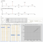
          Multiple HMOs were detected in the urine and plasma of breastfed infants, but not in formula-fed infants. Levels of 2′-fucosyllactose (2′FL), 3FL and lacto-N-neotetraose (LNnT) in both plasma (r = 0.98, p<0.001; r = 0.75, p = 0.002; r = 0.71, p = 0.004) and urine (r = 0.81, p<0.001; r = 0.56, p = 0.026; NS) correlated significantly with concentrations in the corresponding breast milk. The relative fractions of HMOs were low, 0.1% of milk levels for plasma and 4% of milk levels for urine. Within the breastfed cohort, there were significant differences between secretor and nonsecretor groups in levels of several fucosylated HMOs.

          Conclusion

          At least some ingested HMOs are absorbed intact into the circulation and excreted in the urine and their concentrations in these fluids correlate with levels of the corresponding mother's milk. While relative fractions of absorbed HMOs were low, these levels have been shown to have biological effects in vitro, and could explain some of the postulated benefits of human milk.

          Related collections

          Most cited references37

          • Record: found
          • Abstract: found
          • Article: found
          Is Open Access

          Organization of GC/MS and LC/MS metabolomics data into chemical libraries

          Background Metabolomics experiments involve generating and comparing small molecule (metabolite) profiles from complex mixture samples to identify those metabolites that are modulated in altered states (e.g., disease, drug treatment, toxin exposure). One non-targeted metabolomics approach attempts to identify and interrogate all small molecules in a sample using GC or LC separation followed by MS or MSn detection. Analysis of the resulting large, multifaceted data sets to rapidly and accurately identify the metabolites is a challenging task that relies on the availability of chemical libraries of metabolite spectral signatures. A method for analyzing spectrometry data to identify and Qu antify I ndividual C omponents in a S ample, (QUICS), enables generation of chemical library entries from known standards and, importantly, from unknown metabolites present in experimental samples but without a corresponding library entry. This method accounts for all ions in a sample spectrum, performs library matches, and allows review of the data to quality check library entries. The QUICS method identifies ions related to any given metabolite by correlating ion data across the complete set of experimental samples, thus revealing subtle spectral trends that may not be evident when viewing individual samples and are likely to be indicative of the presence of one or more otherwise obscured metabolites. Results LC-MS/MS or GC-MS data from 33 liver samples were analyzed simultaneously which exploited the inherent biological diversity of the samples and the largely non-covariant chemical nature of the metabolites when viewed over multiple samples. Ions were partitioned by both retention time (RT) and covariance which grouped ions from a single common underlying metabolite. This approach benefitted from using mass, time and intensity data in aggregate over the entire sample set to reject outliers and noise thereby producing higher quality chemical identities. The aggregated data was matched to reference chemical libraries to aid in identifying the ion set as a known metabolite or as a new unknown biochemical to be added to the library. Conclusion The QUICS methodology enabled rapid, in-depth evaluation of all possible metabolites (known and unknown) within a set of samples to identify the metabolites and, for those that did not have an entry in the reference library, to create a library entry to identify that metabolite in future studies.
            Bookmark
            • Record: found
            • Abstract: found
            • Article: not found

            Oligosaccharides in human milk: structural, functional, and metabolic aspects.

            Research on human milk oligosaccharides (HMOs) has received much attention in recent years. However, it started about a century ago with the observation that oligosaccharides might be growth factors for a so-called bifidus flora in breast-fed infants and extends to the recent finding of cell adhesion molecules in human milk. The latter are involved in inflammatory events recognizing carbohydrate sequences that also can be found in human milk. The similarities between epithelial cell surface carbohydrates and oligosaccharides in human milk strengthen the idea that specific interactions of those oligosaccharides with pathogenic microorganisms do occur preventing the attachment of microbes to epithelial cells. HMOs may act as soluble receptors for different pathogens, thus increasing the resistance of breast-fed infants. However, we need to know more about the metabolism of oligosaccharides in the gastrointestinal tract. How far are oligosaccharides degraded by intestinal enzymes and does oligosaccharide processing (e.g. degradation, synthesis, and elongation of core structures) occur in intestinal epithelial cells? Further research on HMOs is certainly needed to increase our knowledge of infant nutrition as it is affected by complex oligosaccharides.
              Bookmark
              • Record: found
              • Abstract: found
              • Article: not found

              Physiology of consumption of human milk oligosaccharides by infant gut-associated bifidobacteria.

              The bifidogenic effect of human milk oligosaccharides (HMOs) has long been known, yet the precise mechanism underlying it remains unresolved. Recent studies show that some species/subspecies of Bifidobacterium are equipped with genetic and enzymatic sets dedicated to the utilization of HMOs, and consequently they can grow on HMOs; however, the ability to metabolize HMOs has not been directly linked to the actual metabolic behavior of the bacteria. In this report, we clarify the fate of each HMO during cultivation of infant gut-associated bifidobacteria. Bifidobacterium bifidum JCM1254, Bifidobacterium longum subsp. infantis JCM1222, Bifidobacterium longum subsp. longum JCM1217, and Bifidobacterium breve JCM1192 were selected for this purpose and were grown on HMO media containing a main neutral oligosaccharide fraction. The mono- and oligosaccharides in the spent media were labeled with 2-anthranilic acid, and their concentrations were determined at various incubation times using normal phase high performance liquid chromatography. The results reflect the metabolic abilities of the respective bifidobacteria. B. bifidum used secretory glycosidases to degrade HMOs, whereas B. longum subsp. infantis assimilated all HMOs by incorporating them in their intact forms. B. longum subsp. longum and B. breve consumed lacto-N-tetraose only. Interestingly, B. bifidum left degraded HMO metabolites outside of the cell even when the cells initiate vegetative growth, which indicates that the different species/subspecies can share the produced sugars. The predominance of type 1 chains in HMOs and the preferential use of type 1 HMO by infant gut-associated bifidobacteria suggest the coevolution of the bacteria with humans.
                Bookmark

                Author and article information

                Contributors
                Role: Editor
                Journal
                PLoS One
                PLoS ONE
                plos
                plosone
                PLoS ONE
                Public Library of Science (San Francisco, USA )
                1932-6203
                2014
                7 July 2014
                : 9
                : 7
                : e101692
                Affiliations
                [1 ]Department of Global Discovery Research, Abbott Nutrition, Columbus, Ohio, United States of America
                [2 ]Metabolon, Inc., Durham, North Carolina, United States of America
                Indiana University, United States of America
                Author notes

                Competing Interests: KCG, PAP, and RHB are employed by Abbott Nutrition, whose company funded this study, and own shares of stock in the company. ADK is an employee of Metabolon, Inc. There are no patents, products in development or marketed products to declare. This does not alter the authors' adherence to all the PLOS ONE policies on sharing data and materials.

                Conceived and designed the experiments: KCG RHB. Performed the experiments: ADK. Analyzed the data: KCG ADK. Contributed reagents/materials/analysis tools: KCG ADK RHB. Wrote the paper: KCG ADK PAP RHB.

                Article
                PONE-D-13-46633
                10.1371/journal.pone.0101692
                4085000
                24999728
                1cff0ff2-3f64-4d67-afa2-838c2a685157
                Copyright @ 2014

                This is an open-access article distributed under the terms of the Creative Commons Attribution License, which permits unrestricted use, distribution, and reproduction in any medium, provided the original author and source are credited.

                History
                : 6 November 2013
                : 11 June 2014
                Page count
                Pages: 11
                Funding
                This research was funded by Abbott Nutrition http://abbottnutrition.com/. The funder Abbott Nutrition provided support in the form of salaries for authors KCG, PAP, and RHB, but did not have any additional role in the study design, data collection and analysis, decision to publish, or preparation of the manuscript. The specific roles of these authors are articulated in the ‘author contributions’ section. Metabolon, Inc., provided support in the form of a salary for author ADK, but did not have any additional role in the study design, data collection and analysis, decision to publish, or preparation of the manuscript. The specific roles of these authors are articulated in the ‘author contributions’ section.
                Categories
                Research Article
                Biology and Life Sciences
                Biochemistry
                Metabolism
                Biological Transport
                Glycobiology
                Tissue Distribution
                Immunology
                Clinical Immunology
                Immunomodulation
                Immunity
                Medicine and Health Sciences
                Gastroenterology and Hepatology
                Pediatric Gastroenterology
                Pediatrics
                Neonatology
                Research and Analysis Methods
                Research Design
                Quantitative Analysis

                Uncategorized
                Uncategorized

                Comments

                Comment on this article