9
views
0
recommends
+1 Recommend
0 collections
    0
    shares
      • Record: found
      • Abstract: found
      • Article: found
      Is Open Access

      Frequency-domain vs continuous-wave near-infrared spectroscopy devices: a comparison of clinically viable monitors in controlled hypoxia

      research-article

      Read this article at

      Bookmark
          There is no author summary for this article yet. Authors can add summaries to their articles on ScienceOpen to make them more accessible to a non-specialist audience.

          Abstract

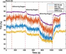
          The Near-infrared spectroscopy (NIRS) has not been adopted as a mainstream monitoring modality in acute neurosurgical care due to concerns about its reliability and consistency. However, improvements in NIRS parameter recovery techniques are now available that may improve the quantitative accuracy of NIRS for this clinical context. Therefore, the aim of this study was to compare the abilities of a continuous-wave (CW) NIRS device with a similarly clinically viable NIRS device utilising a frequency-domain (FD) parameter recovery technique in detecting changes in cerebral tissue saturation during stepwise increases of experimentally induced hypoxia. Nine healthy individuals (6M/3F) underwent a dynamic end-tidal forced manipulation of their expiratory gases to induce a stepwise induced hypoxia. The minimum end-tidal oxygen partial pressure (EtO 2) achieved was 40 mm Hg. Simultaneous neurological and extra-cranial tissue NIRS reading were obtained during this protocol by both tested devices. Both devices detected significant changes in cerebral tissue saturation during the induction of hypoxia (CW 9.8 ± 2.3 %; FD 7.0 ± 3.4 %; Wilcoxon signed rank test P < 0.01 for both devices). No significant difference was observed between the saturation changes observed by either device ( P = 0.625). An observably greater degree of noise was noticed in parameters recovered by the FD device, and both demonstrated equally variable baseline readings (Coefficient of variance 8.4 and 9.7 % for the CW and FD devices, respectively) between individuals tested. No advantageous difference was observed in parameters recovered from the FD device compared with those detected by CW.

          Related collections

          Most cited references17

          • Record: found
          • Abstract: found
          • Article: not found

          Monitoring brain oxygen saturation during coronary bypass surgery: a randomized, prospective study.

          Cerebral deoxygenation is associated with various adverse systemic outcomes. We hypothesized, by using the brain as an index organ, that interventions to improve cerebral oxygenation would have systemic benefits in cardiac surgical patients. Two-hundred coronary artery bypass patients were randomized to either intraoperative cerebral regional oxygen saturation (rSO2) monitoring with active display and treatment intervention protocol (intervention, n = 100), or underwent blinded rSO2 monitoring (control, n = 100). Predefined clinical outcomes were assessed by a blinded observer. Significantly more patients in the control group demonstrated prolonged cerebral desaturation (P = 0.014) and longer duration in the intensive care unit (P = 0.029) versus intervention patients. There was no difference in overall incidence of adverse complications, but significantly more control patients had major organ morbidity or mortality (death, ventilation >48 h, stroke, myocardial infarction, return for re-exploration) versus intervention group patients (P = 0.048). Patients experiencing major organ morbidity or mortality had lower baseline and mean rSO2, more cerebral desaturations and longer lengths of stay in the intensive care unit and postoperative hospitalization, than patients without such complications. There was a significant (r(2) = 0.29) inverse correlation between intraoperative rSO2 and duration of postoperative hospitalization in patients requiring > or =10 days postoperative length of stay. Monitoring cerebral rSO2 in coronary artery bypass patients avoids profound cerebral desaturation and is associated with significantly fewer incidences of major organ dysfunction.
            Bookmark
            • Record: found
            • Abstract: found
            • Article: not found

            Progress of near-infrared spectroscopy and topography for brain and muscle clinical applications.

            This review celebrates the 30th anniversary of the first in vivo near-infrared (NIR) spectroscopy (NIRS) publication, which was authored by Professor Frans Jobsis. At first, NIRS was utilized to experimentally and clinically investigate cerebral oxygenation. Later it was applied to study muscle oxidative metabolism. Since 1993, the discovery that the functional activation of the human cerebral cortex can be explored by NIRS has added a new dimension to the research. To obtain simultaneous multiple and localized information, a further major step forward was achieved by introducing NIR imaging (NIRI) and tomography. This review reports on the progress of the NIRS and NIRI instrumentation for brain and muscle clinical applications 30 years after the discovery of in vivo NIRS. The review summarizes the measurable parameters in relation to the different techniques, the main characteristics of the prototypes under development, and the present commercially available NIRS and NIRI instrumentation. Moreover, it discusses strengths and limitations and gives an outlook into the "bright" future.
              Bookmark
              • Record: found
              • Abstract: found
              • Article: not found

              Review article: cerebral near-infrared spectroscopy in adults: a work in progress.

              Near-infrared spectroscopy (NIRS) has potential as a noninvasive brain monitor across a spectrum of disorders. In the last decade, there has been a rapid expansion of clinical experience using NIRS to monitor cerebral oxygenation, and there is some evidence that NIRS-guided brain protection protocols might lead to a reduction in perioperative neurologic complications after cardiac surgery. However, there are no data to support the wider application of NIRS during routine surgery under general anesthesia, and its application in brain injury, where it might be expected to have a key monitoring role, is undefined. Although increasingly sophisticated apparatuses, including broadband and time-resolved spectroscopy systems, provide insights into the potential of NIRS to measure regional cerebral oxygenation, hemodynamics, and metabolism in real-time, these innovations have yet to translate into effective monitor-guided brain protection treatment strategies. NIRS has many potential advantages over other neuromonitoring techniques, but further investigation and technological advances are necessary before it can be introduced more widely into clinical practice.
                Bookmark

                Author and article information

                Contributors
                0121 371 6741 , David.Davies@uhb.nhs.uk
                Journal
                J Clin Monit Comput
                J Clin Monit Comput
                Journal of Clinical Monitoring and Computing
                Springer Netherlands (Dordrecht )
                1387-1307
                1573-2614
                24 October 2016
                24 October 2016
                2017
                : 31
                : 5
                : 967-974
                Affiliations
                [1 ]ISNI 0000 0004 0376 6589, GRID grid.412563.7, National Institute for Health Research Surgical Reconstruction and Microbiology Research Centre (NIHR SRMRC), , University Hospitals Birmingham NHS Foundation Trust, ; Heritage Building (Old Queen Elizabeth Hospital), Edgbaston, Birmingham, B15 2TH UK
                [2 ]ISNI 0000 0004 0376 6589, GRID grid.412563.7, Department of Neurosurgery, , University Hospitals Birmingham NHS Foundation Trust, ; Birmingham, UK
                [3 ]ISNI 0000 0004 1936 7486, GRID grid.6572.6, PSIBS Doctoral Training Centre, , University of Birmingham, ; Birmingham, UK
                [4 ]ISNI 0000 0004 1936 7486, GRID grid.6572.6, School of Chemistry, , University of Birmingham, ; Birmingham, UK
                [5 ]ISNI 0000 0004 1936 7486, GRID grid.6572.6, School of Sport, Exercise and Rehabilitation Sciences, , University of Birmingham, ; Birmingham, UK
                [6 ]ISNI 0000 0004 1936 7486, GRID grid.6572.6, School of Clinical and Experimental Medicine, , University of Birmingham, ; Birmingham, UK
                Article
                9942
                10.1007/s10877-016-9942-5
                5599440
                27778208
                1c2d232c-b7eb-4985-b546-c9e75238e3ff
                © The Author(s) 2016

                Open AccessThis article is distributed under the terms of the Creative Commons Attribution 4.0 International License ( http://creativecommons.org/licenses/by/4.0/), which permits unrestricted use, distribution, and reproduction in any medium, provided you give appropriate credit to the original author(s) and the source, provide a link to the Creative Commons license, and indicate if changes were made.

                History
                : 29 July 2016
                : 7 October 2016
                Funding
                Funded by: FundRef http://dx.doi.org/10.13039/501100000664, Health Technology Assessment Programme;
                Categories
                Original Research
                Custom metadata
                © Springer Science+Business Media B.V. 2017

                Medicine
                head injury,cerebral blood flow,frequency-domain near-infrared spectroscopy,continuous-wave near-infrared spectroscopy

                Comments

                Comment on this article