7
views
0
recommends
+1 Recommend
1 collections
    0
    shares
      • Record: found
      • Abstract: found
      • Article: found
      Is Open Access

      Clinical Brain Monitoring with Time Domain NIRS: A Review and Future Perspectives

      ,
      Applied Sciences
      MDPI AG

      Read this article at

      ScienceOpenPublisher
      Bookmark
          There is no author summary for this article yet. Authors can add summaries to their articles on ScienceOpen to make them more accessible to a non-specialist audience.

          Abstract

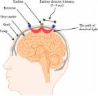
          Near-infrared spectroscopy (NIRS) is an optical technique that can measure brain tissue oxygenation and haemodynamics in real-time and at the patient bedside allowing medical doctors to access important physiological information. However, despite this, the use of NIRS in a clinical environment is hindered due to limitations, such as poor reproducibility, lack of depth sensitivity and poor brain-specificity. Time domain NIRS (or TD-NIRS) can resolve these issues and offer detailed information of the optical properties of the tissue, allowing better physiological information to be retrieved. This is achieved at the cost of increased instrument complexity, operation complexity and price. In this review, we focus on brain monitoring clinical applications of TD-NIRS. A total of 52 publications were identified, spanning the fields of neonatal imaging, stroke assessment, traumatic brain injury (TBI) assessment, brain death assessment, psychiatry, peroperative care, neuronal disorders assessment and communication with patient with locked-in syndrome. In all the publications, the advantages of the TD-NIRS measurement to (1) extract absolute values of haemoglobin concentration and tissue oxygen saturation, (2) assess the reduced scattering coefficient, and (3) separate between extra-cerebral and cerebral tissues, are highlighted; and emphasize the utility of TD-NIRS in a clinical context. In the last sections of this review, we explore the recent developments of TD-NIRS, in terms of instrumentation and methodologies that might impact and broaden its use in the hospital.

          Related collections

          Most cited references125

          • Record: found
          • Abstract: found
          • Article: not found

          A review on continuous wave functional near-infrared spectroscopy and imaging instrumentation and methodology.

          This year marks the 20th anniversary of functional near-infrared spectroscopy and imaging (fNIRS/fNIRI). As the vast majority of commercial instruments developed until now are based on continuous wave technology, the aim of this publication is to review the current state of instrumentation and methodology of continuous wave fNIRI. For this purpose we provide an overview of the commercially available instruments and address instrumental aspects such as light sources, detectors and sensor arrangements. Methodological aspects, algorithms to calculate the concentrations of oxy- and deoxyhemoglobin and approaches for data analysis are also reviewed. From the single-location measurements of the early years, instrumentation has progressed to imaging initially in two dimensions (topography) and then three (tomography). The methods of analysis have also changed tremendously, from the simple modified Beer-Lambert law to sophisticated image reconstruction and data analysis methods used today. Due to these advances, fNIRI has become a modality that is widely used in neuroscience research and several manufacturers provide commercial instrumentation. It seems likely that fNIRI will become a clinical tool in the foreseeable future, which will enable diagnosis in single subjects. Copyright © 2013 Elsevier Inc. All rights reserved.
            Bookmark
            • Record: found
            • Abstract: found
            • Article: not found

            Optical properties of biological tissues: a review.

            A review of reported tissue optical properties summarizes the wavelength-dependent behavior of scattering and absorption. Formulae are presented for generating the optical properties of a generic tissue with variable amounts of absorbing chromophores (blood, water, melanin, fat, yellow pigments) and a variable balance between small-scale scatterers and large-scale scatterers in the ultrastructures of cells and tissues.
              Bookmark
              • Record: found
              • Abstract: found
              • Article: found
              Is Open Access

              fNIRS-based brain-computer interfaces: a review

              A brain-computer interface (BCI) is a communication system that allows the use of brain activity to control computers or other external devices. It can, by bypassing the peripheral nervous system, provide a means of communication for people suffering from severe motor disabilities or in a persistent vegetative state. In this paper, brain-signal generation tasks, noise removal methods, feature extraction/selection schemes, and classification techniques for fNIRS-based BCI are reviewed. The most common brain areas for fNIRS BCI are the primary motor cortex and the prefrontal cortex. In relation to the motor cortex, motor imagery tasks were preferred to motor execution tasks since possible proprioceptive feedback could be avoided. In relation to the prefrontal cortex, fNIRS showed a significant advantage due to no hair in detecting the cognitive tasks like mental arithmetic, music imagery, emotion induction, etc. In removing physiological noise in fNIRS data, band-pass filtering was mostly used. However, more advanced techniques like adaptive filtering, independent component analysis (ICA), multi optodes arrangement, etc. are being pursued to overcome the problem that a band-pass filter cannot be used when both brain and physiological signals occur within a close band. In extracting features related to the desired brain signal, the mean, variance, peak value, slope, skewness, and kurtosis of the noised-removed hemodynamic response were used. For classification, the linear discriminant analysis method provided simple but good performance among others: support vector machine (SVM), hidden Markov model (HMM), artificial neural network, etc. fNIRS will be more widely used to monitor the occurrence of neuro-plasticity after neuro-rehabilitation and neuro-stimulation. Technical breakthroughs in the future are expected via bundled-type probes, hybrid EEG-fNIRS BCI, and through the detection of initial dips.
                Bookmark

                Author and article information

                Contributors
                (View ORCID Profile)
                Journal
                ASPCC7
                Applied Sciences
                Applied Sciences
                MDPI AG
                2076-3417
                April 2019
                April 18 2019
                : 9
                : 8
                : 1612
                Article
                10.3390/app9081612
                81576b1b-67cf-4d7c-8eab-5e5f46c7a804
                © 2019

                https://creativecommons.org/licenses/by/4.0/

                History

                Comments

                Comment on this article