1
views
0
recommends
+1 Recommend
0 collections
    0
    shares
      • Record: found
      • Abstract: found
      • Article: found
      Is Open Access

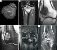
      Images of giant cell tumor and chondroblastoma around the knee: retrospective analysis of 99 cases

      research-article

      Read this article at

      Bookmark
          There is no author summary for this article yet. Authors can add summaries to their articles on ScienceOpen to make them more accessible to a non-specialist audience.

          Abstract

          Background

          It is difficult to differentiate giant cell tumors of the bone (GCTB) from chondroblastoma around the knee based on imaging findings. This study analyzed the imaging features of these 2 diseases for better differentiation.

          Methods

          This retrospective cross-sectional cohort study reviewed data of patients with pathologically confirmed GCTB (n=81; age 15–75 years; median age 33 years) and chondroblastoma (n=18; age 12–34 years; median age 14 years). In all, 18 imaging signs were analyzed.

          Results

          Patients with chondroblastoma were relatively younger than those with GCTB. On imaging, lesion length was significantly (P<0.00001) smaller in chondroblastoma [range, 15.80–78.30 mm; mean ± standard deviation (SD) 34.15±18.24 mm; 95% confidence interval (CI): 24.05–44.25 mm] than in GCTB [range, 30.10–117.50 mm; mean ± SD 59.73±15.28 mm; 95% CI: 56.24–63.22 mm]. Significantly more (P<0.05) chondroblastoma lesions had calcification (76.5% vs. 1.3%), lobulation (77.8% vs. 32.1%), and swelling range >15 mm (84.6% vs. 41.1%) than did GCTB lesions, whereas significantly more (P<0.05) GCTB lesions were greater than half the host bone diameter (74.1% vs. 16.7%) and had a lesion long axis that was consistent with that of the host bone (98.8% vs. 27.8%). There were no significant differences (P>0.05) between the 2 tumors in the remaining 11 imaging signs.

          Conclusions

          A narrow zone of transition, intratumor calcification, lobulation, tumor transverse diameter greater than the bone diameter, maximum lesion length, consistency between the tumor and bone long axes, and edema range around the lesion >15 mm are parameters that can be used to differentiate GCTB from chondroblastoma around the knee.

          Related collections

          Most cited references30

          • Record: found
          • Abstract: found
          • Article: not found

          Giant cell tumor of bone: review, mimics, and new developments in treatment.

          Giant cell tumor (GCT) of bone is generally a benign tumor composed of mononuclear stromal cells and characteristic multinucleated giant cells that exhibit osteoclastic activity. It usually develops in long bones but can occur in unusual locations. The typical appearance is a lytic lesion with a well-defined but nonsclerotic margin that is eccentric in location, extends near the articular surface, and occurs in patients with closed physes. However, GCT may have aggressive features, including cortical expansion or destruction with a soft-tissue component. Fluid-fluid levels, consistent with secondary formation of aneurysmal bone cysts, are seen in 14% of cases. GCT can mimic or be mimicked by other benign or malignant lesions at both radiologic evaluation and histologic analysis. Rarely, GCT is associated with histologically benign lung metastases or undergoes malignant degeneration. In the past, the mainstay of treatment was surgical, primarily consisting of curettage with cement placement, with recurrence rates of 15%-25%. Recurrence is suggested by development of progressive lucency at the cement-bone interface. Other complications include pathologic fracture and postoperative infection. Denosumab, a monoclonal antibody that targets the osteoclastic activity of GCT, has produced 90% tumor necrosis in early studies, results indicative of promise as a potential adjuvant therapy.
            Bookmark
            • Record: found
            • Abstract: found
            • Article: not found

            From the archives of AFIP. Imaging of giant cell tumor and giant cell reparative granuloma of bone: radiologic-pathologic correlation.

            The radiologic features of giant cell tumor (GCT) and giant cell reparative granuloma (GCRG) of bone often strongly suggest the diagnosis and reflect their pathologic appearance. At radiography, GCT often demonstrates a metaepiphyseal location with extension to subchondral bone. GCRG has a similar appearance but most commonly affects the mandible, maxilla, hands, or feet. Computed tomography and magnetic resonance (MR) imaging are helpful in staging lesions, particularly in delineating soft-tissue extension. Cystic (secondary aneurysmal bone cyst) components are reported in 14% of GCTs. However, biopsy must be directed at the solid regions, which harbor diagnostic tissue. These solid components demonstrate low to intermediate signal intensity at T2-weighted MR imaging, a feature that can be helpful in diagnosis. Multiple GCTs, although rare, do occur and may be associated with Paget disease. Malignant GCT accounts for 5%-10% of all GCTs and is usually secondary to previous irradiation of benign GCT. Treatment of GCT usually consists of surgical resection. Recurrence is seen in 2%-25% of cases, and imaging is vital for early detection. Recognition of the spectrum of radiologic appearances of GCT and GCRG is important in allowing prospective diagnosis, guiding therapy, and facilitating early detection of recurrence.
              Bookmark
              • Record: found
              • Abstract: found
              • Article: not found

              Giant cell tumor of bone.

              Giant cell tumor (GCT) of bone is one type of giant cell-rich lesion of bone. This benign mesenchymal tumor has characteristic multinuclear giant cells. Mononuclear stromal cells are the physiologically active and diagnostic cell type. Most GCTs are located in the epiphyseal regions of long bones. The axial skeleton-primarily the sacrum-is a secondary site of involvement. Most patients present with pain, swelling, joint effusion, and disability in the third and fourth decades of life. Imaging studies are important for tumor staging and radiographic grading. Typically, these clinically active but slow-growing tumors are confined to bone, with relatively well-defined radiographic borders. Monostotic disease is most common. Metastatic spread to the lungs is rare. Extended intralesional curettage with or without adjuvant therapy is the primary treatment choice. Local recurrence is seen in ≤ 20% of cases, and a second local intralesional procedure is typically sufficient in cases that are detected early. Medical therapies include diphosphonates and denosumab. Denosumab has been approved for use in osteoporosis as well as breast and prostate cancer metastatic to bone. Medical therapy and radiotherapy can alter the management of GCT of bone, especially in multifocal disease, local recurrences, and bulky central/axial disease.
                Bookmark

                Author and article information

                Journal
                Quant Imaging Med Surg
                Quant Imaging Med Surg
                QIMS
                Quantitative Imaging in Medicine and Surgery
                AME Publishing Company
                2223-4292
                2223-4306
                02 January 2023
                01 February 2023
                : 13
                : 2
                : 787-800
                Affiliations
                [1 ]deptDepartment of Oncology , The Third Hospital of Hebei Medical University , Shijiazhuang, China;
                [2 ]Hebei Provincial Gucheng Hospital, Hengshui , China;
                [3 ]deptDepartment of Radiology , The Third Hospital of Hebei Medical University , Shijiazhuang, China;
                [4 ]deptDepartment of Bone and Soft Tissue Tumors , The Third Hospital of Hebei Medical University , Shijiazhuang, China;
                [5 ]deptDepartment of Medical Research , Shijiazhuang People’s Hospital , Shijiazhuang, China
                Author notes

                Contributions: (I) Conception and design: JL Ma, BH Yu, WJ Wu; (II) Administrative support: WJ Wu; (III) Provision of study materials or patients: JL Ma, BH Yu, Y Wu, C Liu; (IV) Collection and assembly of data: JL Ma, BH Yu, Y Wu, C Liu; (V) Data analysis and interpretation: BH Yu, WJ Wu, BL Gao; (VI) Manuscript writing: All authors; (VII) Final approval of manuscript: All authors.

                [#]

                These authors contributed equally to this work.

                Correspondence to: Zhi-Wei Zhong; Bao-Hai Yu. Department of Radiology, The Third Hospital of Hebei Medical University, 139 Ziqiang Road, Shijiazhuang 050051, China. Email: zhongzhiw@ 123456sina.com ; yubaohai2002@ 123456163.com .
                Article
                qims-13-02-787
                10.21037/qims-22-616
                9929425
                1c20632b-142f-41e9-9baf-fea334415ef2
                2023 Quantitative Imaging in Medicine and Surgery. All rights reserved.

                Open Access Statement: This is an Open Access article distributed in accordance with the Creative Commons Attribution-NonCommercial-NoDerivs 4.0 International License (CC BY-NC-ND 4.0), which permits the non-commercial replication and distribution of the article with the strict proviso that no changes or edits are made and the original work is properly cited (including links to both the formal publication through the relevant DOI and the license). See: https://creativecommons.org/licenses/by-nc-nd/4.0.

                History
                : 16 June 2022
                : 30 November 2022
                Categories
                Original Article

                bone,chondroblastoma,giant cell tumor,imaging,knee
                bone, chondroblastoma, giant cell tumor, imaging, knee

                Comments

                Comment on this article